Research - (2021) Volume 9, Issue 8
Morphometry and Histogenesis of Human Fetal Thymus in Different Gestational Age Groups
*Correspondence: Prabavathy G, Department of Anatomy, Sree Balaji Medical College & Hospital Affiliated to Bharath Institute of Higher Education and Research, India, Email:
Abstract
It was observed that, in one of the cases of 281, the upper pole of the gland appeared as an elongated extension into the root of neck up to the lower border of the thyroid gland and the presence of the lymphocytes from 9th week onwards. formation of lobules had started at 9tli week and distinct formation of lobules were observed at 12th week. Differentiation of the cortex and the medulla became well distinguished from 14th week onwards. the presence of Hassall’s Corpuscles was observed from 14th week and was present in all sections from 15th week onwards and increased in number, size, and maturity with the increase in the gestational age.
Keywords
Corpuscles, Lymphocytes, Thyroid glandIntroduction
Thymus grows rapidly during the embryonic life and childhood, reaches its maximum absolute size about the time of puberty. The thymic components play a responsible role in terminal T-cell differentiation and the development and maintenance of cellular immunity. Knowledge of the embryological, histological, and normal morphological features of the thymus is essential for comprehensive understanding of the normal thymus and thymic diseases [1-3]. Hence this study aims in determining the morphological & histological changes of thymus gland at different gestational weeks in foetus and adults.
Methodology
Thirty-three human foetuses (20 males and 13 females) of different age groups ranging from 9th to 38th gestational week were procured. The Location, extent, shape and the external appearance of the Thymus were observed. Dimensions like Length (1), J3readth (b) & thicknessof the glands were measured using the Digital Vernier Caliper. Furthermore, the tissues obtained were processed and observed.
Results
The collected samples are grouped in to three as group I (9-12 weeks), Group II (13-24 weeks) and group III (25-38 weeks) (Figures 1 to Figure 3).
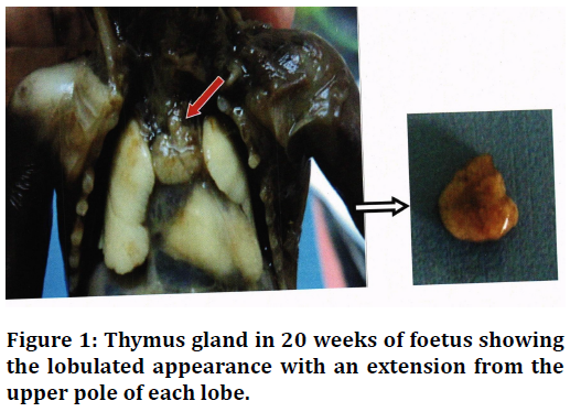
Figure 1. Thymus gland in 20 weeks of foetus showingthe lobulated appearance with an extension from the upper pole of each lobe.
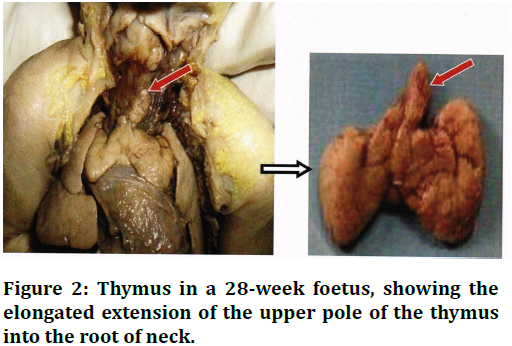
Figure 2. Thymus in a 28-week foetus, showing the elongated extension of the upper pole of the thymus into the root of neck.
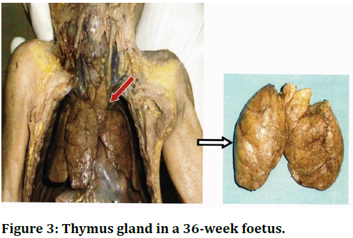
Figure 3. Thymus gland in a 36-week foetus.
Histological studies revealed that the gland was seen to be composed of lymphocytes with a delicate capsule (Figure 4). Human Fetal Thymus (16 weeks) showing the Hassall's Corpuscle in medulla (M) (Figure5). Human Fetal Thymus (34 weeks) showing the Blood vessels in septa (Figure 6).
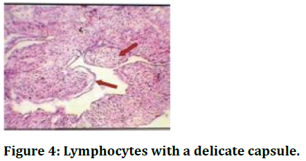
Figure 4. Lymphocytes with a delicate capsule.
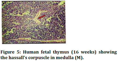
Figure 5. Human fetal thymus (16 weeks) showing the hassall's corpuscle in medulla (M).
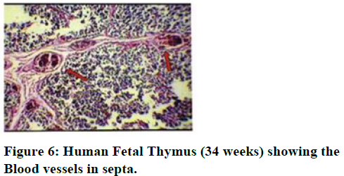
Figure 6. Human Fetal Thymus (34 weeks) showing the Blood vessels in septa.
Discussion and Conclusion
There were different observations regarding the time at which the lymphocytes were present in the thymus such as from 8 weeks onwards. Additional extension from the upper pole of each lobe was observed in most of the foetus of group II and it was well noted in all cases after 3 weeks. there is an increase in length, breath, and thickness of the gland with the increase in gestational age. In a case of adult thymus, histologically distinct lobules were not seen. Differentiation of cortex and medulla also were not distinct [4-7]. Bell et al. [8] reported that the ratio of cortex to medulla decreased with age. The cortex which occupies 60% of the area of section of thymus, decreased to only 30% at 70 years of age. Whereas in the present study, histologically distinct lobules were not seen in the adult thymus. Differentiation of cortex and medulla was also not distinct. Only spare strands of thymic tissue seen within the fatty thymus.
References
- Bodey B, Kaiser HE. Development of Hassall's bodies of the thymus in humans and other vertebrates (especially mammals) under physiological and pathological conditions: immunocytochemical, electronmicroscopic and in vitro observations. In vivo 1997; 11:61-85.
- von Gaudecker B, Schmale EM. Similarities between Hassall's corpuscles of the human thymus and the epidermis. Cell Issue Res 1974; 151:347-68.
- Carlos Ortiz-hidalogo. The man behind hassallâ??s corpuscles. J Clin Pathol 992; 45:99-101.
- Ajita RK, Singh TN, Singh YI, et al. An insight into the structure of the thymus in human foetus-a histological approach. J Anat Soc India 2006; 55:45-9.
- Bratton AB. The normal weight of the human thymus. J Pathol Bacteriol 1925; 28:609-620.
- Davis AE. The histopathological changes in the thymus gland in the acquired immune deficiency syndrome. Annals New York Academy Sci 1984; 437:493-502.
- Haynes BF, Hale LP. The human thymus. Immunol Res 1998; 18:175-192.
- Bell ET. Tumours of the thymus in myasthenia gravis. J Nerve Ment Dis1917; 45: 130.
Author Info
Department of Anatomy, Sree Balaji Medical College & Hospital Affiliated to Bharath Institute of Higher Education and Research, Chennai, Tamil Nadu, IndiaCitation: Prabavathy G,Morphometry and Histogenesis of Human Fetal Thymus in Different Gestational Age Groups, J Res Med Dent Sci, 2021, 9(8): 62-63
Received: 14-Jul-2021 Accepted: 02-Aug-2021
