Research - (2021) Volume 9, Issue 6
Natural Compounds AS D2 Receptor Agonist, M4 Receptor Antagonist and ACHE Modulator: Mechanistic and in Silico Modelling Studies
M Ganga Raju*, Veena Yadav and NVL Suvarchala Reddy V
*Correspondence: M Ganga Raju, Department of Pharmacology, Gokaraju Rangaraju College of Pharmacy, Bachupally, Hyderabad 500090, Telangana, India, Email:
Abstract
Catalepsy is a behavioural phenomenon where the rodent is not able to change an externally imposed posture, and antipsychotic-induced catalepsy is a rodent model for the motoric side effects often seen when using antipsychotic medication. The present study is focused on the evaluation of methonolic extract of Tagetus patula as anti-catalepsy against haloperidol (1.0 mg/kg, i.p) and Reserpine (5 mg/kg, i.p,) induced catalepsy in mice using levodopa as standard. The biochemical parameters like reduced glutathione (GSH) and catalase were estimated in mice brain in reserpine induced catalepsy. The extract was screened against lipid peroxidation assay and DPPH radical scavenging assay using ascorbic acid as standard. The cataleptic score in haloperidol and reserpine treated groups were found to be higher. But in groups treated with the METP and standard drug (levodopa 25 mg/kg, i.p) the cataleptic score was found to be reduced in both the models. Upon treatment with extract at 200 mg/kg. bd. wt, 400 mg/kg. bd. wt. & levodopa 25 mg/kg, i.p, significant improvement in GSH and catalase levels were observed. The results clearly showed the ability of the extract to scavenge the free radicals in both the assays. In the present study docking studies were performed for natural compounds present in the extract such as Patuletin, Quercetin, δ-elemene, Tagetenone, β-Ocimene, Piperitenone, Tagetone, Limonene, β-ionone, β-caryophyllene and standard drug Levodopa against PDB ID: 3INJ, PDB ID: 1DSG, PDB ID: 6EP4 using schrodinger software. The statistical verification of the model was evaluated with PROCHEK; a structure verification program relies on Ramachandran plot which determines the quality of the predicted structures. The results revealed that Patuletin, Quercetin, Piperitenone, Limonene and δ-elemene had shown highest glide scores which indicates a stronger receptor-ligand binding affinity among the various phytochemical constituents present in the extract. According to Ramachandran plot a good model would be expected to have over 90% in most favoured regions. In our study all the selected protein models have more than 90% favored regions clearly indicating that the selected models are of good quality. From the above it is concluded that the extract possesses anti-cataleptic and anti-oxidant activity.
Keywords
Levodopa, Tagetus patula, Docking studies, Ascorbic acid, Schrodinger software.
Introduction
The etiology of catalepsy is a state characterized by muscle rigidity and failure to correct an externally imposed posture for a prolonged period. In addition, catalepsy induced by the typical neuroleptic agent haloperidol in rodents can be used as a model of the extrapyramidal effects often seen in Parkinson’s disease [1] and resulted in the generation of significant oxidative stress in their brain regions, as evidenced by the increase of the lipid peroxidation product, malonaldehyde and catecholamine metabolism by monoamine oxidases (MAOs). Haloperidol causes movement disorders such as the narcoleptics malignant syndrome, dystonia’s and tardive dyskinaesias [2]. Reserpine 4–5 mg/kg, s.c.) work by inhibiting the vesicular monoamine transporter, VMAT2. This leads to loss of storage capacity and hence depletion of brain (and peripheral) monoamines including noradrenaline and 5- HT as well as dopamine. Oxidative stress, which is a culprit in many human diseases, has been implicated in haloperidol toxicity. The catalepsy test is widely used to evaluate motor effects of drugs that act on the extra pyramidal system. Evidence suggests that immense oxidative stress, free radical formation, genetic susceptibility, and programmed cell death are the main causes for neurodegeneration associated with Parkinson’s and other related diseases [3]. Levodopa is used as standard drug in anticataleptic activity.
Tagetes patula, commonly called French marigold of family (Asteraceae). It is native to South America, where it has culinary and medical uses. Different parts of plants are used to color human foods, animal-based textiles, for antifungal activity, anti-arthritic, immunomodulatory and anti-inflammation [4-6]. The present study aimed to evaluate the neurobehavioral and neuroprotective effect of the extract of Tagetes patula on Reserpine- induced and haloperidol-induced catalepsy in mice.
Materials and Methods
Plant collection and drying
Flowers of Tagetes patula were identified, collected, authenticated by botanist Rabiya Sultana, New government degree college, Kukatpally. Tagetes patula flowers were cleaned and dried under shade for about six days and powdered. The powdered material was stored.
Preparation of methanolic extract of Tagetes patula (Soxhlet)
The powdered material of flower heads of Tagetes patula were dried and extracted with methanol by soxhlation technique. As to get efficient extraction, this method allows a continuous extraction process; it is nothing but a series of short macerations. The organic extracts obtained were evaporated to dryness by keeping at room temperature. Large amounts of drug can be extracted with a much smaller quantity of solvent. This process of extraction is economical in terms of time, energy and consequently financial investments [7].
Preliminary phytochemical analysis of the extract
The extract was subjected to preliminary phytochemical investigations to identify various phytoconstituents present in the methanolic extract of flower heads of Tagetes patula.
Acute toxicity testing
The acute toxicity studies were carried out using OECD 425 guidelines. Present study was carried out in CPCSEA approved animal house of Gokaraju Rangaraju College of Pharmacy, Bachupally, Hyderabad, India. (Reg.No. 1175/PO/ERe/S/08/CPCSEA).
Animal housing
The animals (mice) were housed in poly acrylic cages with not more than six animals per cage, with 12 h light/12 h dark cycle. Animals have free access to standard diet and drinking water ad libitum. The animals were allowed to acclimatize the laboratory environment for a week before the start of the experiment. The care and maintenance of the animals were carried out as per the approved guidelines of the committee for the purpose of control and supervision of experiments on animals (CPCSEA).
In vivo methods for evaluation of anticataleptic activity
In vivo evaluation of anticataleptic activity of the methanolic extract of flower heads of Tagetes patula was carried out in following models.
Haloperidol induced catalepsy.
Reserpine induced catalepsy.
Haloperidol induced catalepsy
30 Swiss albino mice of either sex weighing about 25-30 gm were selected for this study. They were divided into five groups with 6 animals in each group. Group I: normal control receives normal saline. Group II: disease control receives Haloperidol (1.0 mg/kg, i.p). Group III, IV, receives test drug METP at dose of 200 mg/k and 400 mg/kg. Group V receives (standard) levodopa 25 mg/kg, i.p. Except group I, all the remaining groups receives haloperidol daily for a period of 7 days to induce catalepsy. On the last day after 30 min of the administration of drugs, the animals were subjected to standard bar test. Severity of catalepsy was measured for every 30 min, thereafter up to a total duration of 2 hours. Catalepsy of individual mice was measured in a stepwise manner by a scoring method in stages as described below. The method assessed the ability of an animal respond to an externally imposed posture.
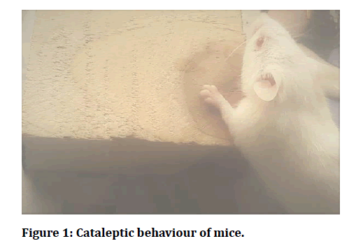
Stage I: The mice was taken out of the home cage and placed on a table. If mice move freely no score was given. Stage II: If the mice failed to move when touched gently on the back or pushed, score of 0.5 was assigned. Stage III: The front paws of the mice were placed alternately on a 3 cm high block. If the mice failed to correct the posture within 15 sec, a score of 0.5 for each paw was added to the score of step I. Stage IV: The front paws of the mice were placed alternately on a 6 cm high block. If the mice failed to correct the posture within 15 sec, a score of 1 for each paw was added to the scores of steps I and step II. Thus, for an animal, the highest score was 3.5 (cut-off score) and that reflects total catalepsy [8] (Figure 1).
Figure 1: Cataleptic behaviour of mice.
Reserpine induced catalepsy
30 Swiss albino mice of either sex weighing about 25-30 gm were selected for this study. They were divided into five groups with 6 animals in each group. Group I: normal control receives normal saline. Group II: disease control receives reserpine (5 mg/kg, i.p). Group III, IV, receives test drug METP at dose of 200 mg/k and 400 mg/kg. Group V receives (standard) levodopa 25 mg/kg, i.p. Except group I, all the remaining groups receives reserpine on alternative days for a period of 5 days to induce catalepsy. On the 5th day catalepsy was measured by using standard bar test. After completion of the behavioural study animals were euthanized and the brain was isolated and homogenized. The homogenate was used for estimation of biochemical parameters GSH and catalase [9].
Biochemical estimations
Estimation of catalase
The catalase activity assay was carried out as described by Beers and Sizer. The reaction mixture consists of 2 ml phosphate buffer (pH 7.0), 0.95 ml of hydrogen peroxide (0.019 M) and 0.05 ml supernatant in final volume of 3 ml. Absorbance was recorded at 240 nm. One unit of catalase was defined as the amount of enzyme required to decompose 1 m mol of peroxide per minute, at 25°C and at pH 7.0. The results were expressed as units of catalase activity milligram per protein [10].
Estimation of reduced glutathione (GSH)
The amount of GSH in mice brain was measured according to the method of Sedlak et al. 1968). Briefly, brain tissue was deproteinized with an equal volume of 10% trichloroacetic acid and was allowed to stand at 40°C for 2 h. The contents were centrifuged for 15 min. The supernatant was added to tris buffer (pH 8.9) containing ethylene diamine tetra acetic acid (EDTA) (pH 8.9) followed by the addition of 0.01 M 5, 5, 0-dithiobis (2-nitrobenzoic acid) (DTNB). Finally, the mixture was diluted with distilled water, to make the total mixture to 3 ml and absorbance was read in a spectrophotometer at 412 nm and results were expressed as l g GSH/g tissue [9].
In vitro antioxidant assay
In vitro evaluation of antioxidant activity of the methanolic extract of flower heads of Tagetes patula was carried out in following models.
Lipid peroxidation assay
Lipid peroxide formation was measured by lipid peroxidation assay by the modified method of Ohkawa et al. and Masao et al. Mice (weighing 25-30 g) were sacrificed by dislocation of the neck. The brain was removed and then homogenized in phosphate buffer saline (pH 7.0). 1 ml of brain homogenate (10%, w/v) was added to the test extract of different concentrations. The lipid peroxidation was initiated by adding 100 μl of 15 mM FeSO4 solution. After 30 min of incubation at room temperature, 0.1 ml of reaction mixture (brain homogenate + test drug) was taken in a tube containing 0.1 ml of SDS (8.1%w/v), 0.75 ml of 20% trichloroacetic acid and 0.75 ml of 0.8% TBA solution. The volume in each tube was made up to 2 ml with distilled water and then heated on water bath at 95°C for 60 minutes. After 60 minutes, the volume in each tube was made up to 2.5 ml and then 2.5 ml of N butanol: pyridine (5:1) was added in each tube. The reaction mixture was vortexed and centrifuged at 4000 rpm for 10 minutes. The organic layer was removed and absorbance was read at 532 nm in a UV spectrophotometer. The experiment was performed in triplicate. The percentage inhibition was calculated using the formula [11].
Percentage inhibition=Abscontrol–Abssample × 100/ (Abcontrol)
DPPH radical scavenging assay
The DPPH assay is popular in natural product antioxidant studies, hydrogen donor is an antioxidant that measures compounds that are radical scavengers. DPPH is one of the few stable and commercially available organic nitrogen radicals. The antioxidant effect is proportional to the disappearance of DPPH in test samples. Monitoring DPPH with a UV spectrometer has become the most used method because of its simplicity and accuracy. DPPH shows a strong absorption maximum at 517 nm (purple). The color turns from purple to yellow followed by the formation of DPPH upon absorption of hydrogen from an antioxidant. This reaction is stoichiometric with respect to the number of hydrogen atoms absorbed. Therefore, the antioxidant effect can be easily evaluated by following the decrease of UV absorption at 517 nm.
To METP 5 ml of 0.004% (w/v) solution of DPPH was added. The obtained mixture was vortexed, incubated for 30 min in room temperature in a relatively dark place and then was read using spectrophotometer at 517 nm. The blank was 80% (v/v) methanol. Ascorbic acid (vitamin C) was used for comparison. Measurements were taken in triplicate. DPPH scavenging effect was calculated using the following equation:
DPPH Percentage inhibition (%)=A0–A × 100 /(A0)
Where A0 is the absorbance of negative control (0.004% DPPH solution) and A is the absorbance in presence of extract. The results were reported as IC50 values and ascorbic acid equivalents (AAE, mg/g) of METP [12].
Molecular docking studies
Selection of proteins: PDB ID: 3INJ, 1DSG, 6EP4
Many antipsychotic compounds, e.g., haloperidol and risperidone at high doses, frequently induce extrapyramidal side effects in patients [13] an effect most likely caused by dopamine D2 receptor blockade [14]. The cataleptic effects of dopamine D2 receptor antagonists may be caused by increased release of striatal acetylcholine that subsequently binds to muscarinic receptors located on medium spiny striatopallidal or striatonigral neurons. Muscarinic receptor agonists worsen haloperidol-induced catalepsy whereas muscarinic receptor antagonists exhibit the opposite effect [15]. The precise mechanism of this action is unknown. The muscarinic M4 receptor would be an obvious target, due to its pronounced localization in the striatum [16]. The cataleptic effect of antipsychotics is triggered by blockade of D2R on cholinergic interneurons and the consequent increase of acetylcholine signaling on striatal projection neurons.
These studies illuminate the critical role of D2Rmediated signaling in regulating the activity of striatal cholinergic interneurons and the mechanisms of typical antipsychotic side effects. So, AChE agonist would also be an obvious target in the treatment of catalepsy induced by antipsychotic drugs. The present study focused on the interactions of D2 receptor agonist, (PDB ID: 3INJ), M4 receptor antagonist (PDB ID: 1DSG) and AchE agonist (PDB ID: 6EP4) with natural compounds present in the extract and the standard drug Levodopa using Schro. dinger software.
Statistical analysis
Values are expressed as Mean ± SEM, (n=6). All the groups were compared with control, negative control, and standard by using Dunnett’s t-test. Significant values are expressed as control group (**=p<0.01, *=p<0.05), negative control (A=p<0.01, B=p<0.05) and standard (a=p< 0.01, b=p< 0.05), ns- nonsignificant.
Results
Methanolic extract of flower heads of Tagetes patula was explored for its in vivo anticataleptic activity usingsuitable rodent models and antioxidant activity using in vitro models. All the results obtained in the study were included below.
Preparation of methanolic extract of flower heads of Tagetes patula
The methanolic extract of flower heads of Tagetes patula was prepared by soxhlation technique. The percentage yield of methanolic extract was calculated by using the following formula.
% of yield obtained=Amount of extract obtained (gms)/ Total amount powder used × 100
% Yield of extract=45/500 ×100=9% w/w
Preliminary phytochemical analysis
The preliminary phytochemical investigation of methanolic extract of flower heads of Tagetes patula revealed the presence of bioactive compounds of which phenolic acids, flavonoids, essential oils, thiophene derivatives, benzofunan derivatives, alkaloids, tannins were the most prominent (Table 1).
| Phytochemical constituents | Result |
|---|---|
| Phenolic acids | ++ |
| Flavonoids | ++ |
| Essential oils | ++ |
| Thiophene derivatives | ++ |
| Benzafunan derivatives | ++ |
| Carotenoids | + |
| Alkaloids | + |
| Tannins | + |
Note: + indicates present
Table 1: Preliminary phytochemical analysis.
Acute toxicity studies
Methanolic extract of flower heads of Tagetes patula was tested on swiss albino mice up to a dose of 2000 mg/kg bd. wt. The animal did not exhibit any signs of toxicity or mortality up to 2000 mg/kg bd. wt. various morphological and behavioral characters were observed during the study. Hence the extract was found to be safe up to 2000 mg/kg bd. wt.
Dose selection
From toxicity studies, a dose of 2000 mg/kg bd. wt. was identified to be safe, and the working dose was considered as 1/10th i.e., 200 mg/kg. bd. wt. In the present study pharmacological evaluations were done using 200 mg/kg. bd. wt and 400 mg/kg. bd. wt.
In vivo anti cataleptic activity
The methanolic extract of flower heads of Tagetes patula was screened for its anticataleptic activity using the following models.
Haloperidol induced catalepsy
From table 2 the cataleptic score in haloperidol treated group was found to be higher. But in groups treated with the METP and standard drug (levodopa 25 mg/kg, i.p) the cataleptic score was found to be reduced.
| Groups | Treatment | Cataleptic score | |||
|---|---|---|---|---|---|
| 30 min | 60 min | 90 min | 120 min | ||
| I | Saline water (p.o) | 0 | 0 | 0 | 0 |
| II | Haloperidol (1 mg/kg, i.p) | 2.88 ± 0.22**a | 3 ± 0.16**a | 3.25 ± 0.15**a | 3.33 ± 0.09**a |
| III | METP (200 mg/kg, p.o) | 2.58 ± 0.21*bA | 2.33 ± 0.19**aA | 2.16 ± 0.15**bB | 2 ± 0.26**bA |
| IV | METP (400 mg/kg, p.o) | 1.83 ± 0.19**bA | 1.66 ± 0.19**b A | 1.41 ± 0.14*bA | 1.16 ± 0.09**A |
| V | Levodopa (25 mg/kg, i.p) | 1.33 ± 0.25*A | 1.16 ± 0.15*A | 1 ± 0.11*A | 0.83 ± 0.15*A |
Values are expressed as Mean ± SEM, (n=6). Statistical analysis was performed by using ANOVA followed by Dunnett’s test. Results were compared with control group (**p<0.01, *p<0.05), disease control (A = p<0.01, B = p<0.05) and standard (a = p<0.01, b = p<0.05).
Table 2: Effect of METP on haloperidol induced catalepsy (standard bar test).
Reserpine induced catalepsy
From table 3 in METP and levodopa treated groups the cataleptic score was found to be reduced when compared to negative control group. The cataleptic score in reserpine treated group was found to be higher.
| Groups | Treatment | Cataleptic score | |||
|---|---|---|---|---|---|
| 30 min | 60 min | 90 min | 120 min | ||
| I | Saline water (p.o) | 0 | 0 | 0 | 0 |
| II | Reserpine (5 mg/kg, i.p) | 3.08 ± 0.14**a | 3.25 ± 0.15**a | 3.41 ± 0.07**a | 3.5 ± 0**, a |
| III | METP (200 mg/kg, p.o) | 2.75 ± 0.19**aA | 2.75 ± 0.19**aB | 2.3 ± 0.19**aA | 2.08 ± 0.21**a |
| IV | METP (400 mg/kg, p.o) | 2.08 ± 0.21**A | 1.83 ± 0.21**A | 1.5 ± 0.21**bA | 1.33 ± 0.19**bA |
| V | Levodopa (25 mg/kg, i.p) | 1.5 ± 0.26*bA | 1.33 ± 0.25**A | 1.08 ± 0.26**A | 0.91 ± 0.14**A |
Values are expressed as Mean ± SEM, (n=6). Statistical analysis was performed by using ANOVA followed by Dunnett’s t-test. Results were compared with control group (*= p<0.01, **=p<0.05), disease control (A=p<0.01, B=p<0.05) and standard (a= p<0.01, b=p<0.05).
Table 3: Effect of METP on reserpine induced catalepsy (standard bar test).
Effect of METP on catalase and GSH in reserpine induced catalepsy model
The effect of METP on biochemical parameters (catalase and GSH) are tabulated in the table 4. As the pathogenesis of the catalepsy is attributed to the increase in the oxidative stress which in turn is associated with the antioxidant enzymes so the effect of METP wasevaluated on the levels of enzymes like catalase and GSH. From the results the enzymes like catalase and GSH were found to be lowered in reserpine treated group. But the content of these two enzymes were found to be improved in extract and standard drug treated groups.
| Group | Treatment | Catalase (n mol H2O2/mg protein) | Reduced glutathione (GSH) (μg/mg protein) |
|---|---|---|---|
| I | Saline water (p.o) | 7.5 ± 0.43 | 17.1 ± 0.34 |
| II | Reserpine (5 mg/kg, i.p) | 2.85 ± 0.19**, a, A | 6.42 ± 0.41**, a, A |
| III | METP (200 mg/kg, p.o) | 4.9 ± 0.3*, b, A | 14.55 ± 056*, b, A |
| IV | METP (400 mg/kg, p.o) | 5.21 ± 0.13*, b, A | 15.8 ± 0.50*, b, A |
| V | Levodopa (25 mg/kg, i.p) | 6.86 ± 0.14**, A | 16.21 ± 0.74**, A |
Values are expressed as Mean ± SEM, (n=6). Statistical analysis was performed by using ANOVA followed by Dunnett’s t-test. Results were compared with control group (* = p < 0.01, **=p<0.05), disease control (A=p<0.01) and standard (a=p<0.01, b=p<0.05).
Table 4: Effect of METP on catalase and GSH.
In vitro antioxidant assay
The methanolic extract of flower heads of Tagetes patula was subjected to in vitro antioxidant activity. In vitro antioxidant activity was performed using.
Lipid peroxidation assay
DPPH assay.
Lipid peroxidation assay
In vitro antioxidant activity was performed using lipid peroxidation assay. The results were expressed in table 5.
| S. No | Compounds | Concentration (µg/ml) | Percentage inhibition | IC50 value |
|---|---|---|---|---|
| 1 | METP | 10 | 18.03 ± 0.33 | 31 |
| 20 | 30.7 ± 0.33 | |||
| 30 | 57.9 ± 0.86 | |||
| 40 | 68.2 ± 0.68 | |||
| 50 | 79.2 ± 1.60 | |||
| 2 | Ascorbic Acid | 10 | 14.2 ± 0.25 | 26 |
| 20 | 26.8 ± 0.31 | |||
| 30 | 50.2 ± 0.89 | |||
| 40 | 63.5 ± 0.30 | |||
| 50 | 73.03 ± 0.20 |
Table 5: Lipid peroxidation assay of methanolic extract of flower heads of Tagetes patula.
In lipid peroxidation assay, METP was tested at different concentrations like 10, 20, 30, 40, and 50 μg/ml. Percentage inhibition and IC50 values are tabulated in the table 5. In recent years lipid peroxidation, is a crucialstep in the pathogenesis of several disease states.(Hussein RH et al., 2014). The reducing property of METP indicates that they can be used as electron donors who reduced the oxidized intermediates of lipid peroxidation process; therefore, it can be used as an antioxidant.
DPPH radical scavenging assay
In vitro antioxidant activity was performed using DPPH radical scavenging assay. The results were expressed in table 6.
| S. No | Compounds | Concentration (µg/ml) | Percentage inhibition | IC50 value |
|---|---|---|---|---|
| 1 | METP | 10 | 20.9 ± 0.38 | 30 |
| 20 | 32.6 ± 0.32 | |||
| 30 | 58.4 ± 2.3 | |||
| 40 | 67.3 ± 0.83 | |||
| 50 | 76.06 ± 0.75 | |||
| 2 | Ascorbic acid | 10 | 17.5 ± 0.84 | 27 |
| 20 | 26.5 ± 0.75 | |||
| 30 | 49.7 ± 0.50 | |||
| 40 | 64.7 ± 0.38 | |||
| 50 | 71.6 ± 0.94 |
Table 6: DPPH radical scavenging assay of methanolic flower extract of flower heads of Tagetes patula.
In DPPH radical scavenging assay, METP was tested at different concentrations like 10, 20, 30, 40, and 50 μg/ mland percentage inhibition and IC50 values are tabulated in the table 6. The IC50 value for the METP was found to be 30 μg/ml which is compared with standard ascorbic acid having IC50 value of 27 μg/ml respectively.
Molecular docking against
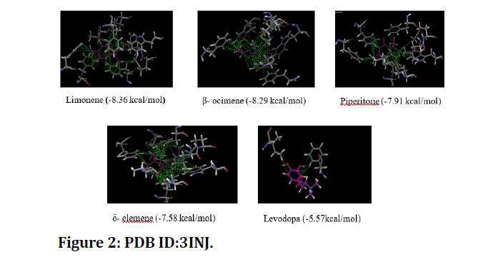
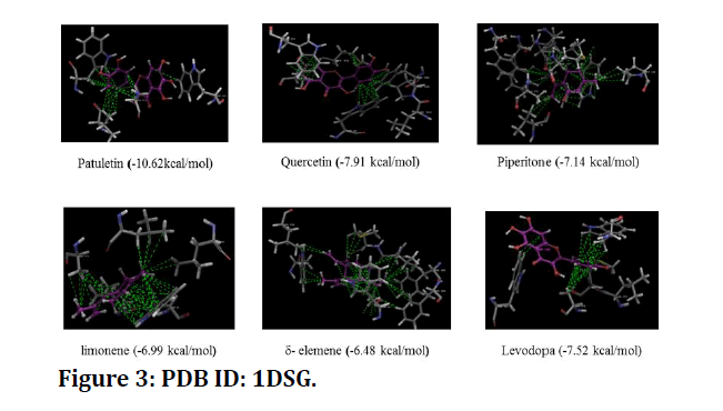
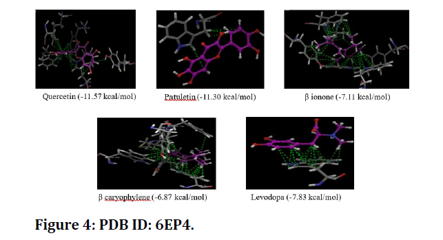
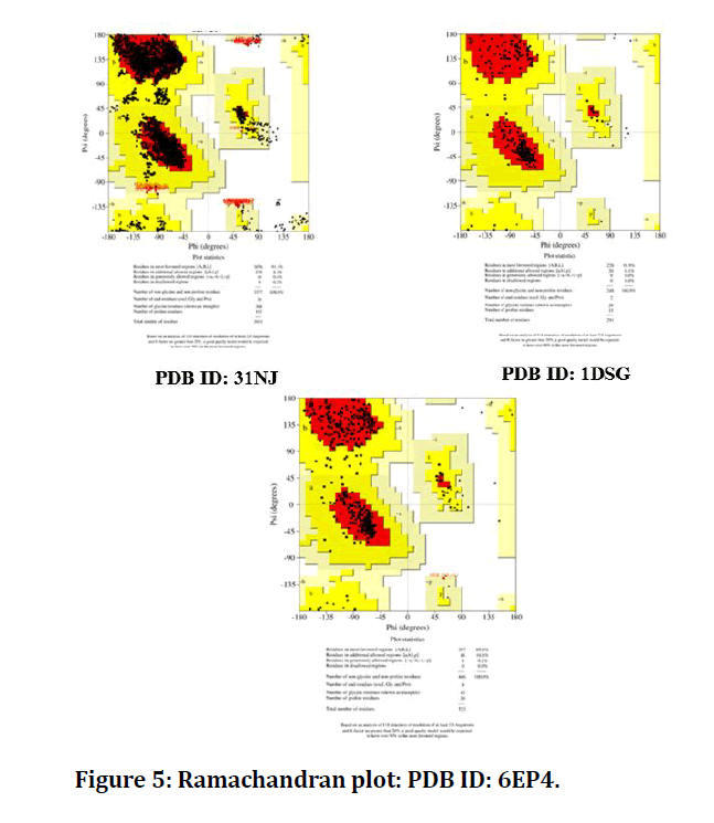
Results are explained in the form of figure (Figure 2 to Figure 4) and tables (Table 7). Ramachandran plot explained in Figure 5.
Figure 2: PDB ID:3INJ.
Figure 3: PDB ID: 1DSG.
Figure 4: PDB ID: 6EP4.
Figure 5: Ramachandran plot: PDB ID: 6EP4.
| Constituent | Docking Score (kcal/mol) | ||
|---|---|---|---|
| PDB ID: 3INJ | PDB ID: 1DSG | PDB ID: 6EP4 | |
| Patuletin | -6.62 | -10.62 | -11.3 |
| Quercetin | -6.65 | -7.91 | -11.57 |
| Delta elemene | -7.58 | -6.48 | -6.44 |
| Tagetenone | -7.11 | -6.37 | -6.59 |
| Beta Ocimene | -8.29 | -5.77 | -6.21 |
| Piperitenone | -7.91 | -7.14 | -- |
| Tagetone | -6.11 | -6.08 | -- |
| Limonene | -8.36 | -6.99 | -- |
| Beta ionone | -- | -5.62 | -7.11 |
| Beta caryophyllene | -- | -6.31 | -6.87 |
| Levodopa | -5.57 | -7.52 | -7.83 |
Table 7: Glide score of proteins by molecular docking (Schrodinger software).
Discussion
Anti-cataleptic and antioxidant activity
The present study revealed the anti-cataleptic effect of methanolic extract of flower heads of Tagetes patula in haloperidol and reserpine induced catalepsy and antioxidant activity against lipid peroxidation and DPPH radical scavenging assay. Catalepsy is a condition in which the animal maintains imposed posture for long time before regaining the normal posture. Haloperidol is a typical antipsychotic drug (a non-selective D1 and D2 dopamine antagonist) induced catalepsy in the striatum. Reserpine induces catalepsy by interfering with the storage of catecholamines in intracellular granules, resulting in monoamine depletion (norepinephrine, 5- hydroxytryptamine and dopamine) in nerve terminals and induces hypolocomotion and muscular rigidity. In our study the methanolic flower head extract of Tagetes patula at two dose levels showed significant reduction in the cataleptic scores in both haloperidol and reserpine induced catalepsy. A Reduction in the cataleptic score is an indicative of anti-cataleptic activity. Levodopa act as a precursor for the dopamine when administered it crosses the blood brain barrier and is up taken by the surviving dopaminergic neurons, converted to dopamine which is stored and released as a transmitter [17].
The literature reveals the presence of various phytochemical constituents like flavonoids, phenolic acids, essential oils, thiophene derivatives, alkaloids, carotenoids, and benzafunan derivatives in Tagetes patula [18,19]. Flavonoids found to activate the endogenous antioxidant levels in neuronal cells hence protecting them from undergoing neurodegeneration [20]. The flavonoids like quercetagetin, 6- hydroxykaempferol, phenols, carotenoids like lutein, linoliec acid and essential oils were reported to possess antioxidant activity [21] by upregulating the production of intracellular antioxidant enzymes such as SOD, GPx, CAT and glutathione in neuroleptics induced catalepsy [22,23]. Beside this, quercetin was also found to protect neuronal cells from oxidative stress through elevation of intracellular glutathione levels [24]. Flavonoids and polyphenols are also known to act through prevention of chain initiation, decomposition of peroxides, reducing capacity and radical scavenging. Terpenes (limonene) have a wide range of biological properties ranging from antioxidant, anti-inflammation, and anti-cancer to neuroprotection [25,26].
Limonene (+) acts as an antioxidant, its activity being the potential main mechanism contributing to the beneficial effect of limonene (+) on human health. Indeed, an in vitro study showed protective effects of limonene (+) on H2O2-induced chromosome breakage and loss along with DNA damage on human lymphocytes and V79 cell lines [27]. Furthermore, in a number of other studies, limonene (+) has been shown to be beneficial to health by inhibiting oxidative stress [28,29].
It is also well established that the administration of haloperidol leads to an increase in the oxidative stress in the brain tissue [30]. Under normal conditions, decreased activity of antioxidant enzymes, such as SOD, glutathione peroxidase and catalase, in the brain leads to the accumulation of oxidative free radicals resulting in degenerative effects [31]. An increase in these enzymes under normal conditions would represent increased antioxidant activity and a protective mechanism in neuronal tissue, thus, constituting the first line of defense against oxidative stress in our body. So, any overall decrease in cataleptic scores and SOD activity in the drug treated groups indicates the ability of the drug extract to combat oxidative stress in brain tissue and reduce the severity of haloperidol-induced catalepsy. The various chemical constituents present in the METP might be responsible for its anticataleptic activity and antioxidant activity.
Molecular docking:
Molecular docking continues to hold great promise in the field of computer based drug design which screens small molecules by orienting and scoring them in the binding site of a protein. The docking analysis of isolated compounds from methanolic extract of Tagetes patula and standard drug like levodopa were carried out using schrodinger software. The various constituents identified in the plant extract are Patuletin, Quercetin, δ-elemene, Tagetenone, β-Ocimene, Piperitenone, Tagetone, Limonene, β-ionone, β-caryophyllene and standard drug Levodopa are subjected to docking against PDB ID: 3INJ, PDB ID: 1DSG, PDB ID: 6EP4. The proteins identified namely PDB ID: 31NJ, PDB ID: 1DSG, PDB ID: 6EP4 are modeled and the qualities of the 3D model were evaluated using the PROCHECK program and assessed using the Ramachandran plot. It is evident from the Ramachandran plot that predicted models have most favorable regions, additionally allowed regions, generally allowed regions and disallowed regions. Such a percentage distribution of the protein residues determined by Ramachandran plot shows that the predicted models are of good quality. According to Ramachandran plot a good quality model would be expected to have over 90 % in most favored regions.
The results revealed that Patuletin, quercetin, piperetone, limonene and β-ocimene showed highest glide scores among the various phytochemical constituents present in the extract against all the protein models selected. β- ocimene, limonene, piperotone, tagetenone, δ-elemene, showed highest glide score against PDB, ID: 31NJ. Patuletin, quercetin, piperitone and levodopa have shown high score against PDB ID: 1DSG where as Patuletin, quercetin, beta-ionone and levodopa against PDB ID: 6EPA.
The glide scores of some of the constituents namely patuletin, quercetin, and β- ocimene present in the extract were found to be higher than the standard drug levodopa stating that the compounds have more affinity to bind to the proteins. These results clearly indicate that the chemical constituents might have shown similar mechanism of action to that of the standard drug levodopa in reducing the catalytic score.
Conclusion
The methanolic extract of flower heads of Tagetes patula possess anti-cataleptic and antioxidant activity in rodent models. Further studies are needed to be carried out to isolate individual phytochemical constituents of the extract and to establish the exact mechanism for its anticataleptic and antioxidant activities.
References
- Dhanalakshmi S, Khaserao SS, Kasture SB. Effect of ethanolic extract of some anti- asthmatic herbs on clonidine and haloperidol- induced catalepsy in mice. OPEM 2004; 4:1-5.
- Jayachandra P, Prabhakaran V, Umasankar K, et al. Anti-cataleptic activity of ethanol extract of Vernonia cinerea. Asian J Pharm Sci Technol 2012; 2:23-29.
- Rasheed A, Venkataraman S, Fazil A, et al. Evaluation of antioxidant potential of Smilax zeylanica linn. In reversing haloperidol induced catalepsy in rats. Int J Pharm Pharm Sci 2012; 4.
- Yasukawa K, Kasahara, Y. Effects of flavonoids from french marigold (Florets of Tagetes patula L) on acute inflammation model. Int J Inflamm 2013; 1-5.
- Jabeen A, Mesaik M, Simjee S, et al. Anti-TNF- α and anti-arthritic effect of patuletin: A rare flavonoid from Tagetes patula. Int Immunopharmacol 2016; 36:232-240.
- Kashif M, Bano S, Naqvi S, et al. Cytotoxic and antioxidant properties of phenolic compounds from Tagetespatula flower, Pharmac Biol 2015; 53:672-681.
- Raju MG, Srilakshmi S, Reddy NVLS. Anti-inflammatory, in-silico docking and ADME analysis of some isolated compounds of Tagetes erecta flower heads. Int J Pharmac Sci Res 2020; 11:1358-1366.
- Jayachandra P, Prabhakaran V, Umasankar K, et al. Anti-cataleptic activity of ethanol extract of Vernonia cinerea. Asian J Pharm Sci Technol 2017; 2.
- Vishakha T, Rachana D, Siddhi M, et al. Evaluation of neuroprotective activity of bauhinia variegata on reserpine induced catalepsy in rats. J Med Sci Clin Res 2015; 3:7421-7428.
- Souravh B, NS Gill, Nitan K. Neuroprotective effect of juniperus communis on chlorpromazine induced parkinson disease in animal model. Chinese J Biol 2015; 1-7.
- Sakat SS, Juvekar A R, Gambhire MN. In vitro antioxidant and anti-infammatory activity of methanol extract of oxalis corniculata Linn. Int J Pharm Pharmaceut Sci 2010; 2:146–155.
- Mahdi-Pour B, Jothy S, Latha L, et al. Antioxidant activity of methanol extracts of different parts of Lantana camara. Asian Pacific J Tropical Biomed 2012; 2:960-965.
- Gao K, David KE, Stephen GJ, et al. Antipsychotic-induced extrapyramidal side effects in bipolar disorder and schizophrenia. J Clin Psychopharmacol 2008; 28:203-209.
- Arnt J, Skarsfeldt T. Do novel antipsychotics have similar pharmacological characteristics? A review of the evidence. Neuropsychopharmacol 1998; 18:63–101.
- Klemm WR. Evidence for a cholinergic role in haloperidol-induced catalepsy. Psychopharmacology 1985; 85:139–142.
- Hersch SM, Gutekunst CA, Rees HD, et al. Distribution of M1-M4 muscarinic receptor proteins in the rat striatum-light and electron-microscopic immunocytochemistry using subtype-specific antibodies. J Neurosci 1994; 14:3351–3363.
- Tripathi KD. Essentials of medical pharmacology. 7th Edn. Jaypee Brothers Medical Publishers (P) Ltd, New Delhi, 2013; 816-835.
- Faizi S, Naz A. A novel and labile β-carboline alkaloid from the flowers of Tagetes patula. Tetrahedron 2002; 58:6185-6197.
- Magalingam KB, Radhakrishnan AK, Nagaraja H. Protective mechanisms of flavonoids in parkison’s disease. Oxidative Med Cellular Longevity 2015; 1-14.
- XU Li-wei, Chen J, Huan Y, et al. Phytochemical and their biological activities in Tagetes L. Chinese Herbal Med 2012; 4:103-117.
- Wiegand H, Wagner AE, Boesch-Saadatmandi C, et al. Effect of dietary genistein on Phase II and antioxidant enzymes in rat liver. Cancer Genomics Proteomics 2009; 6:85-92.
- Floyd RA, Carney JM. “Free radical damage to protein and DNA: Mechanisms involved and relevant observations on brain undergoing oxidatives tress. Annals Neurol 1992; 32:S22–S27.
- Ishige K, Schubert D, Sagara Y. Flavonoids protect neuronal cells from oxidative stress by three distinct mechanisms. Free Radical Biol Med 2001; 30:433–446.
- Cho KS, Lim YR, Lee K, et al. Terpenes from forests and human health. Toxicol Res 2017; 33:97-106.
- Trombetta D, Castelli F, Sarpietro MG, et al. Mechanisms of antibacterial action of three monoterpenes. Antimicrob Agents Chemotherapy 2005; 49:2474-2478.
- Bacanlı M, BaÅ?aran AA, BaÅ?aran N. The antioxidant and antigenotoxic properties of citrus phenolics limonene and naringin. Food Chem Toxicol 2015; 81:160-170.
- Bai J, Zheng Y, Wang G, et al. Protective effect of δ-limonene against oxidative stress-induced cell damage in human lens epithelial cells via the p38 pathway. Oxidative Med Cellular Longevity 2016; 2016:5962832.
- Suh KS, Chon S, Choi EM. Limonene protects osteoblasts against methylglyoxalderived adduct formation by regulating glyoxalase, oxidative stress, and mitochondrial function. Chem Biol Interactions 2017; 278:15-21.
- Sagara Y. Induction of reactive oxygen species in neurons by haloperidol. J Neurochem 1998; 71:1002–1012.
- Naidu PS, Singh A, Kulkarni SK. Effect of Withania somnifera root extract on haloperidol induced orofacial dyskinesia: Possible mechanism of action. J Med Food 2003; 6:107–114.
- Aragno M, Brignardello E, Tamagno O, et al. Dehydroeppiandrosterone administration prevents the oxidative damage induced by acute hyperglycemia in rats. J Endocrinol 1997; 155:233–240.
Author Info
M Ganga Raju*, Veena Yadav and NVL Suvarchala Reddy V
Department of Pharmacology, Gokaraju Rangaraju College of Pharmacy, Bachupally, Hyderabad 500090, Telangana, IndiaCitation: M Ganga Raju, Veena Yadav, NVL Suvarchala Reddy V, Natural Compounds AS D2 Receptor Agonist, M4 Receptor Antagonist and ACHE Modulator: Mechanistic and in Silico Modelling Studies, J Res Med Dent Sci, 2021, 9(6): 1-6
Received: 12-Apr-2021 Accepted: 09-Jun-2021 Published: 17-Jun-2021

