Research Article - (2022) Volume 10, Issue 8
Optical Biosensor for Detection of Rheumatoid Arthritis via Cadherin-11
Alaa Mohsen Ali* and Layla MH Al-ameri
*Correspondence: Alaa Mohsen Ali, Department of Oral Medicine, University of Baghdad, Institute of Laser for Postgraduate Studies, Bag, Iraq, Email:
Abstract
Optical biosensors exhibit various analytical techniques because they enable direct, accurate, and label-free detection of a wide range of biological and chemical substances. Rheumatoid Arthritis (RA) is an autoimmune disease that can cause joint pain and damage throughout the body. The joint damage that RA causes usually happens on both sides of the body. So, if a joint in one arm or leg is affected, the same joint in the other arm or leg will probably be affected too.
Aim: The aim of this study is using a new method for detection Cadherin-11 in serum by optical biosensor instead of detection it in serum by using Elisa technique in conventional method for diagnosis of Rheumatoid arthritis.
Materials and Methods: By using Elisa technique, the concentration of CDH11 is determined after treatment with chemical drugs (MTX).Then these samples of patients examined optically by laser biosensor which constructed from a source of light (diode of 405 nm) connected to multimode-no core-multimode fibre which are connected to ocean (HR2000) spectrum analyser by an adapter and to the PC for display the results.
Results: A change in the wavelength and intensity of transmitted light have been recorded with the change of the concentration of CDH11 after treatment with MTX (chemical therapy).
Conclusion: We could conclude from this study that the optical biosensor is suitable method for accurate, rapid detection of CDH11 and its concentration in rheumatoid arthritis patients' rather than a conventional method which use Elisa technique.
Keywords: Optical biosensor, Cadherin-11 (CDH11), Optical fibre, Rheumatoid arthritis
Introduction
Rheumatoid Arthritis (RA) is an autoimmune and inflammatory disease with chronic systemic characterized by synovitis and vasculitis that causes cartilage destruction, joint distortion, loss of joint function, and systemic organ damage, affecting approximately 0.5-1% of the population [1]. The synovium is affected by the infection caused by rheumatoid arthritis. By producing synovial fluid, this membrane lines and lubricates the joints, resulting in synovitis, which induces joint pain, stiffness, and swelling [2]. In RA patients, the minor joints (wrists, elbows, knees, ankles, and hands and feet) are most commonly affected by inflammation. However, any joint in the body could be harmed. While RA generally affects the joints, synovitis can broaden and damage other organs and tissues, making RA a systemic disease [3].
Cadherin’s are calcium dependent haemophilic cell to cell adhesion Trans membrane proteins. Cadherin’s cytoplasmic tail binds to catenin, which connects the cadherin to the actin cytoskeleton. Cadherin’s regulate cellular behaviour in ways other than adhesion. Cadherin’s, in particular, are essential regulators of cell migration and invasion. Cadherin’s have also been linked to the regulation of the Epithelial to Mesenchyme Transition (EMT) and the differentiation of my fibroblasts [4]. Cadherin-11 (CDH11) is a calcium-dependent, type II traditional cadherin that was originally described on osteoblasts but is now represented by fibroblasts, my fibroblasts, injured type II alveolar epithelial cells, and macrophages [5]. The entire Cadherin-11 gene is expressed in various tissues, including the heart, brain, placenta, lung, bone, also involved in the development of joint formation, both physically and pathologically [6].
According to that, several studies have been done about cadherin-11 for different disease. Mesas Pedro et al. discovered that targeting Cadherin-11 reduces skin fibrosis in the tight skin-1 mouse model [4]. Daniele Vergers et al. developed an SPR based immunoassay for the sensitive detection of the soluble epithelial marker E-cadherin [7]. Young-Eon Park, et al. experimentally modelled the IL-17 increases Cadherin-11 for arthritis and in rheumatoid arthritis detection [8]. Mestas Pedro et al. investigated the role of Cadherin-11 in liver fibrosis caused by carbon tetrachloride [9].
Biosensors are devices that detect the concentrations of biological substances and convert them to electrical signals [10]. Biosensors are analytical tools that include biologically sensitive materials immobilized as recognition elements (such as enzymes, antibodies, antigens, microorganisms, cells, tissue, nucleic acid, and other biologically active substances), physicochemical transducers, and other biologically active substances (such as electrochemical electrodes, photodiodes, and signal-amplifying devices) [11,12]. Laser biosensor architecture components are less expensive, reliable, and precise than test strips. SPR, waveguides, optical fibre, and other laser based detection methods are used in various biosensors [13]. Laser based biosensors are essential in many fields, such as immunoassays and drug screening, due to their high sensitivity and accuracy [14]. Optical fibres could be used to miniaturize the most critical laser based detection approach used in many instruments [15]. In biosensors, laser detection based on transient waves is commonly used. In recent years, laser biosensors have proven to be handy tools in various fields, including pharmaceutical research, analytical biochemistry, food environmental experiments, and diagnostic methods [16]. Biosensors are used in applications such as disease observation, drug detection, pollutant detection, disease by microorganisms, and biomarkers, which are disease markers in body fluids (urine, saliva, blood, sweat) [17].
There is no simple technique for detecting and evaluating rheumatoid arthritis in its early stages (RA). The aim of this study is to determine CDH11 for diagnostic purposes and determine its concentration by using optical biosensor; instead of conventional technique (Elisa); which offers rapid and more accurate results for RA.
Materials and Methods
Sample collection: Ten millilitres of blood were drawn from each patient's, and then centrifuged to obtain serum, and this serum was divided into two parts, each one of them was five millilitres in size. The first part was used to manually detect of Cadherin-11 (CDH11) using an ELISA technique, the other part (5 ml) is used for detection the Cadherin-11 (CDH11) by using optical biosensor.
Biological measurement: The Elisa technique has been used to assay CDH11 level in human serum. The sandwich-Elisa method is used in this technique where the micro ELISA strip plate has been pre-coated with a CDH11 antibody and the standards or samples are placed in the appropriate micro ELISA strip plate wells and mixed with the appropriate antibody. Then, in each Micro Elisa strip plate well, a Horse Radish Peroxidase (HRP)-conjugated antibody specific for CDH11 is added and incubated. Other elements have been washed away. Each well receives the TMB substrate solution. Only the wells containing CDH11 and HRP-conjugated CDH11 antibodies will appear blue before turning yellow when the stopping solution is added. At 405 nm, the Optical Density (OD) is measured spectrophotometric-ally. The OD value is proportional to CDH11 concentration. Comparing the OD of the sample to the standard curve could calculate the concentration of CDH11 in the samples.
Laser biosensor set-up structure
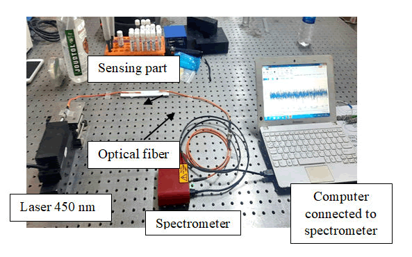
The multimode optical fibre with a 30 cm length has been cut (from the middle) to produce two fragment of multimode fibre with 15 cm of each one and about 2 cm of coreless optical fibre insert between them, then a groove was made on both sides. The buffer part was removed from the optical fibre by immersing it in 40% acetone for 30 minutes and then washing it with distilled water to remove all impurities and thoroughly cleaning it. In 10 minutes, a portion of the fibre is immersed in pure Hydrofluoric acid (HF) 40% to remove the fibre’s cladding, and then it is washing with distilled water. The entire fibre (30 cm) was inserted into the aplastic device using an adhesive silicone. The fibres’ ends are connected to a transformer device. Optical fibre connectors connect the terminal tools to the optical fibres. Splicing connected two organized fibres to form a continuous optical waveguide. The laser exporter (a blue diode laser) will be connected to the first end of the fibre, which is connected to a power supply. A diode laser with a 450 nm wavelength and an output power has been used. This laser wavelength has been chosen depending on the absorption spectra of the protein, where it matched the absorption peaks of the protein Cadherin-11, where it is measured by using a spectrophotometer. The other side has been connected to a spectrum analyser (ocean HR2000) to measure the intensity of signal. The optical spectrum analyser (Ambient Optics HR2000) was then connected to the spectrometer (via a USB port to a computer), with an accuracy of 0.035 nm at a high wavelength and working in the 200-1100 nm wavelength range (Figure 1).
Figure 1: Set up of biosensor.
Results and Discussion
Measurement the concentration of CDH11
A serum sample for 7 patients (male and female) for measuring the concentration of CDH11 after different treatment (chemical and biological) by conventional method and optically detection by biosensor to detect CDH11 and determine its concentration spectrally via the intensity and wavelength of transmitted light. Table 1 show the concentration of CDH11 after chemo (MTX) treatment measured by Elisa technique.
| No. | CDH11 N.V (6–200 pg/ml | Treatment (chemical, biological) | Gender | Age |
|---|---|---|---|---|
| 1 | 22.7 Normal | MTX | M | 16 |
| 2 | 28.8 Normal | MTX | F | 33 |
| 3 | 280↑ | MTX | F | 15 |
| 4 | 307.5↑ | MTX | F | 28 |
| 5 | 512.6↑ | MTX | M | 16 |
| 6 | 618.2↑ | MTX | M | 22 |
| 7 | 790↑ | MTX | F | 10 |
Table 1: The concentrations of CDH11 after chemical treatment.
The statistical analysis of these results show a significant value of CDH11 after treatment (MTX) in Table 2
| Treatment | Cadherin frequency (%) | Probability | |||
|---|---|---|---|---|---|
| Males | Females | ||||
| Normal | High | Normal | High | ||
| MTX | 1 (50.0) | 2 (100.0) | 3 (75.0) | 1 (11.1) | P>0.05 |
Table 2: Statistical analysis of CDH11 MTX treatments.
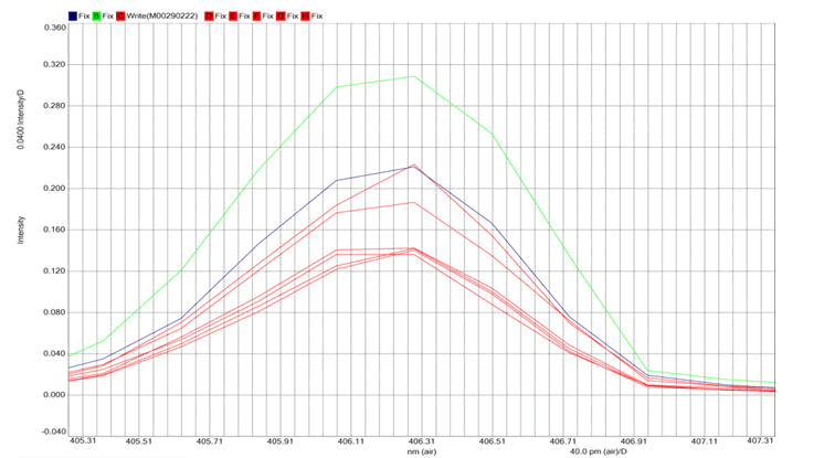
By laser biosensor, the CDH11 is detected and the concentration of CDH11 is determined by through the change in the intensity of transmitted light and shifting of the transmitted wavelength with the change of the concentration of CDH11 after treatment with MTX (chemical therapy) as the optical spectrum analyser is shown in Figure 2 where it represent the intensity of transmitted light as a function of the transmitted wavelength with respect to the concentration of CDH11. Where the spectrum of CDH11 is determine at its standard and normal level as a result of matching its peak absorption with the source of light wavelength, and then the change of the transmitted intensities and shifting of the wavelength were got as the concentration of CDH11 increase. Where with the increasing of the CDH11 concentration, the absorption of light will be increased and the intensity of transmitted light will be decreased while the wavelength of the transmitted light will be increased. So the optical biosensor succeeds in detecting CDH11 and its concentration with very high accuracy for diagnostic purposes of rheumatoid arthritis.
Figure 2: Intensity of transmitted light as a function of transmitted wavelength with respect to the concentration of CDH11.
Green line (source); blue line (normal concentration) Red line (different concentration of CDH11)
Conclusion
In the present paper an optical biosensor have been used for detection on rheumatoid arthritis by detecting on CDH11 in blood serum for the patient of rheumatoid arthritis. An MM-no core- MM fibre sensor and the laser source (405 nm) detect Cadherin-11 (CDH11) levels. The biosensor gave an excellent result for the examined serum where it was succeed in detecting for CDH11. According to the study's findings, the highest laser intensity corresponds to the lowest protein concentration and vice versa. Finally, we conclude that the optical biosensor is a more effective, more stable, sensitive, with a rapid diagnosis and cheaper method for detecting CDH11 more than the traditional method.
Reference
- Ma D, Liang N, Zhang L. "Establishing Classification Tree Models in Rheumatoid Arthritis Using Combination of Matrix-Assisted Laser Desorption/Ionization Time-of-Flight Mass Spectrometry and Magnetic Beads." Front Med 2021; 8:1–12.
- Driver CB, Stoppler MC, "Rheumatoid Arthritis (RA): Early Signs," Symptoms, Causes, Treat. Diet 2020.
- Imas JJ, Zamarreno CR, Zubiate P, et al. Optical biosensors for the detection of rheumatoid arthritis (Ra) biomarkers: A comprehensive review. Sensors 2020; 20:1–51.
[Crossref] [Google Scholar][Pubmed]
- Pedroza M, Welschhans RL, Agarwal SK. Targeting of cadherin-11 decreases skin fibrosis in the tight skin-1 mouse model. PLoS One 2017; 12:1–9.
[Crossref] [Google Scholar][Pubmed]
- Dou C, Yan Y, Dong S. Role of cadherin-11 in synovial joint formation and rheumatoid arthritis pathology. Mod Rheumatol 2013; 23:1037–1044.
[Crossref] [Google Scholar][Pubmed]
- Manning JE, Lewis JW, Marsh LJ, et al. Insights into Leukocyte Trafficking in Inflammatory Arthritis–Imaging the Joint. Front Cell Dev Biol 2021; 9:1–4.
- Vergara D, Bianco M, Pagano R, et al. An SPR based immunoassay for the sensitive detection of the soluble epithelial marker E-cadherin. Nanomedicine 2018; 14:1963–1971.
[Crossref] [Google Scholar][Pubmed]
- Park YE, Woo YJ, Park SH, et al. IL-17 increases cadherin-11 expression in a model of autoimmune experimental arthritis and in rheumatoid arthritis. Immunol Lett 2000; 140: 97–103.
[Crossref] [Google Scholar][Pubmed]
- Pedroza M, To S, Smith J, et al. Cadherin-11 contributes to liver fibrosis induced by carbon tetrachloride. PLoS One 2019; 14:1–11.
[Crossref] [Google Scholar][Pubmed]
- Damborsky P, Svitel J, KatrlÃk J. Optical biosensors. Essays Biochem 2016; 60:91–100.
[Crossref] [Google Scholar][Pubmed]
- Correia R, James S, Lee SW, et al. Biomedical application of optical fibre sensors. J Opt 2018; 20:1–26.
[Crossref] [Google Scholar][Pubmed]
- Abed HASL, Alameri LM. Detection of the Level of Human Alt Liver Enzyme Concentration By Using Laser Biosensor Multimode Fibres (Mmf). Biochem Cell Arch 2019; 19:3829–3833.
- Shatti R, Al-ameri LMH. Optically Sensing for Thyroid Profile Hormones in Blood. Med Leg Updat 202121:172–176.
- Mohammed SE, Al-ameri LMH. Laser Bio stimulation Effect on Human Sperm Motility. Iraqi J Laser 2021; 20:39–42.
- Salman NA, Taher HJ, Mohammed SA. Tapered Splicing Points SMF-PCF-SMF Structure based on MachZehnder interferometer for Enhanced Refractive Index Sensing. Iraqi J Laser 2017; 16:19-24.
- Al-Ameri LMH, Faris RA, Belal SJ, "Detection of HbA1c in Blood Using Diode Laser (491) nm," Syst Rev Pharm 2021; 12:700–704.
- Addanki S, Amiri IS, Yupapin P. "Review of optical fibres-introduction and applications in fibre lasers." Results Phys 2018; 10:743–750.
Author Info
Alaa Mohsen Ali* and Layla MH Al-ameri
Department of Oral Medicine, University of Baghdad, Institute of Laser for Postgraduate Studies, Bag, IraqCitation: Alaa Mohsen Ali, Layla MH Al-ameri, Optical Biosensor for Detection of Rheumatoid Arthritis via Cadherin-11, J Res Med Dent Sci, 2022, 10 (8): 000-000.
Received: 03-Jun-2022, Manuscript No. JRMDS-22-57153; , Pre QC No. JRMDS-22-57153; Editor assigned: 07-Jun-2022, Pre QC No. JRMDS-22-57153; Reviewed: 21-Jun-2022, QC No. JRMDS-22-57153; Revised: 04-Aug-2022, Manuscript No. JRMDS-22-57153; Published: 16-Aug-2022


