Research - (2023) Volume 11, Issue 1
PREVALENCE OF ACCESSORY CANALS IN PRIMARY MANDIBULAR MOLARS-A RADIOGRAPHIC ANALYSIS
Cibikkarthik T and Mahesh Ramakrishnan*
*Correspondence: Mahesh Ramakrishnan, Department of Pedodontics, Saveetha Dental College and Hospitals, Saveetha Institute of Medical and Technical Sciences, Saveetha University, Tamil Nadu, India, Email:
Abstract
Objective: Detailed anatomy of any canal configuration forms the base of successful endodontic treatment. The study planned to evaluate the accessory canals in primary mandibular molars Materials and methods: Eight hundred cases were obtained from a central database of clinical management software in a private Dental University from March 2020 to March 2021.SPSS were used for all the statistical test. Results: Results from the study revealed that most of the accessory canals are seen higher among the male population 57.52% when compared with female population 42.48%. Most of the accessory canals are seen in 2-7 years of age 79.17% and in 2nd mandibular molars 3 canals were common. Conclusion: Predominantly there were three canals in males compared to females. But the difference was not statistically significant. Almost three or four canal configurations are the common variant seen.
Keywords
Primary molars, Accessory canal, Root and canal morphology, Innovative technique
Introduction
Canal configuration is the most important criteria for successful endodontic treatment in childrens [1]. Through understanding of the variations by the clinicians helps to eliminate bacteria in the canal and proper biomechanical preparation [2–6]. The canal configuration is more complex in primary molars compared to that of permanent molars and also presence of various resorption patterns can also affect the success rate of the procedure.
In the primary dentition the mandibular arch seems to be affected more when compared to maxillary arch, while maxillary incisors are more to carious, the molars are more commonly affected in the mandibular. This can be attributed to the fact that mandibular molars exhibit deeper pit san fissures [7]. High sugar diet and improper feeding habits combined with difficulty in maintaining good oral hygiene plays an important role in carie formation in children, the enamel is also less mineralized in children which can results in early involvement of pulp [8]. When there is caries it results in secondary and tertiary dentin secretion and various rsportion patterns which can alter the number of root canals present at every stage. Radiographs may tend to show a different canal pattern but histological sectioning is a must to understand the through pattern [9].
The furcation area of a molar tooth, which encompasses part around the division of roots is most important in a primary molars because of presence of multiple accessory canals and also the permanent tooth germ is formed below the furcation area. Hence any changes in the furcation area can involve the permanent tooth which can even lead to enamel deformation. Turner hypoplasia is the most common pathology which is an enamel defect caused due to periapical infection in the primary molars [10].
Advances in imaging modalities such as use of CBCT in identifying canal morphology of complex cases has a disadvantage of high radiation dose. The children when exposed to higher dose of radiation can exhibit adverse effects; hence the concept of ALARA should be weighed before any imaging. Most of the clinical situations IOPA remain the most common imaging technique due to less exposure and convenient in children [11]. Most of our team members have published high quality epidemiology and various researches [12–26]. This study was performed to determine the prevalence of accessory canals in the primary mandibular molars.
Materials and Methods
Study setting
It was carried out as a retrospective study in a private Dental University in Southern Part of India. The advantage is the study population belongs to a particular ethnic group and the disadvantage is it’s a single centric study. A total of 86000 case records were screened by trained reviewers and entered in excel sheets. .calibration of the examiners was done using a pilot study and kappa statistics were done. The number of roots, age and gender were entered in the tabular column.
Results and Discussion
Out of total sample size (725 cases), results from the study reveals that occurrence of accessory canals in mandibular molars was higher among the male population 57.52% when compared with female population 42.48% (Figure 1); 79.17% of accessory canals are seen in the age of 2-7 years and 20.83% in 8-12 years (Figure 2); 57.93% of the accessory canals are seen in the mandibular 2nd molars and 42.07% in mandibular 1st molars (Figure 3). In mandibular molars 52.28% of 3 canals are commonly seen and 37.66% of 4 canals and 10.07% of 5 canals (Figure 4). Further assessment of the prevalence of accessory canals revealed that 27.45% of 3 canals, 23.31% of 4 canals and 7.17% of 5 canals seen in 2nd mandibular molars. The correlation between the number of canals and the mandibular region revealed that Pearson Chi-Square Value-0.001;(p<0.05). Hence statistically significant (Figure 5).
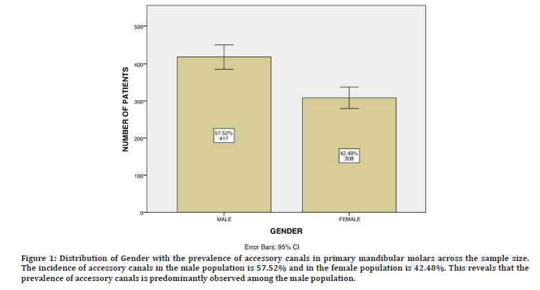
Figure 1: Distribution of Gender with the prevalence of accessory canals in primary mandibular molars across the sample size. The incidence of accessory canals in the male population is 57.52% and in the female population is 42.48%. This reveals that the prevalence of accessory canals is predominantly observed among the male population.
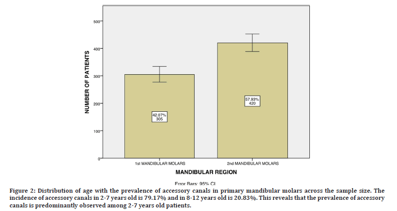
Figure 2: Distribution of age with the prevalence of accessory canals in primary mandibular molars across the sample size. The incidence of accessory canals in 2-7 years old is 79.17% and in 8-12 years old is 20.83%. This reveals that the prevalence of accessory canals is predominantly observed among 2-7 years old patients.
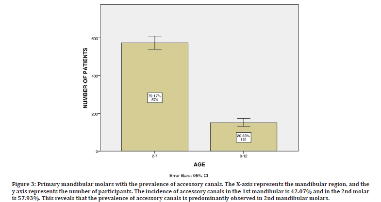
Figure 3: Primary mandibular molars with the prevalence of accessory canals. The X-axis represents the mandibular region, and the y axis represents the number of participants. The incidence of accessory canals in the 1st mandibular is 42.07% and in the 2nd molar is 57.93%. This reveals that the prevalence of accessory canals is predominantly observed in 2nd mandibular molars.
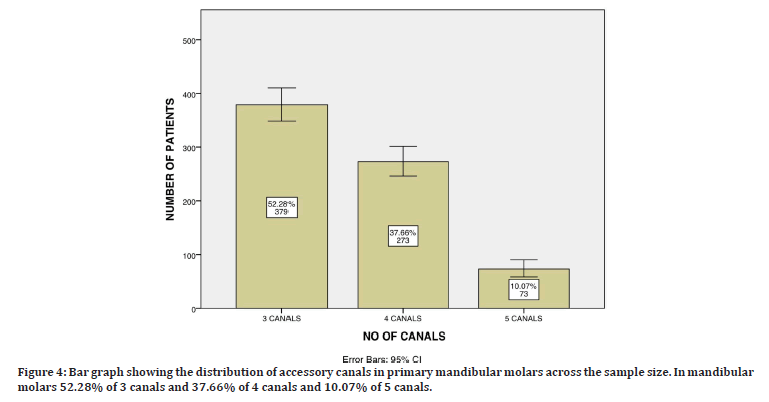
Figure 4: Bar graph showing the distribution of accessory canals in primary mandibular molars across the sample size. In mandibular molars 52.28% of 3 canals and 37.66% of 4 canals and 10.07% of 5 canals.
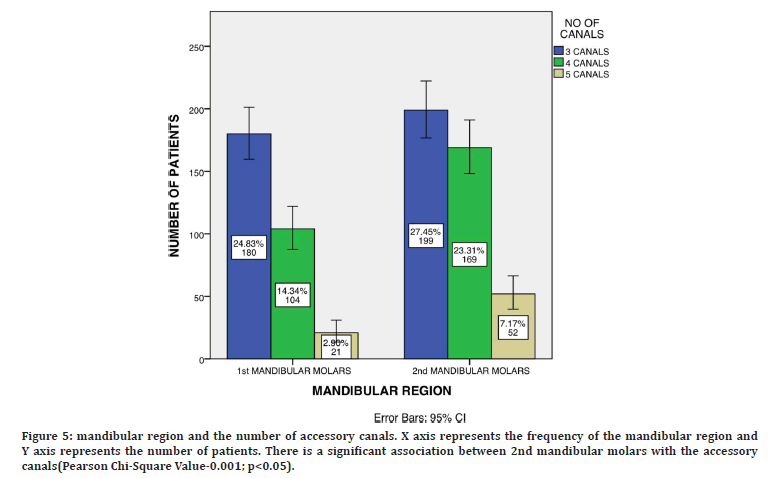
Figure 5: mandibular region and the number of accessory canals. X axis represents the frequency of the mandibular region and Y axis represents the number of patients. There is a significant association between 2nd mandibular molars with the accessory canals(Pearson Chi-Square Value-0.001; p<0.05).
An assessment of the gender with the prevalence of accessory canals revealed that 30.34% of 3 canals, 21.93% of 4 canals and 5.24% of 5 canals were seen in male population. The correlation between the gender and the prevalence of accessory canals revealed that Pearson Chi-Square Value-0.307;p>0.05. Hence statistically not significant (Figure 6).
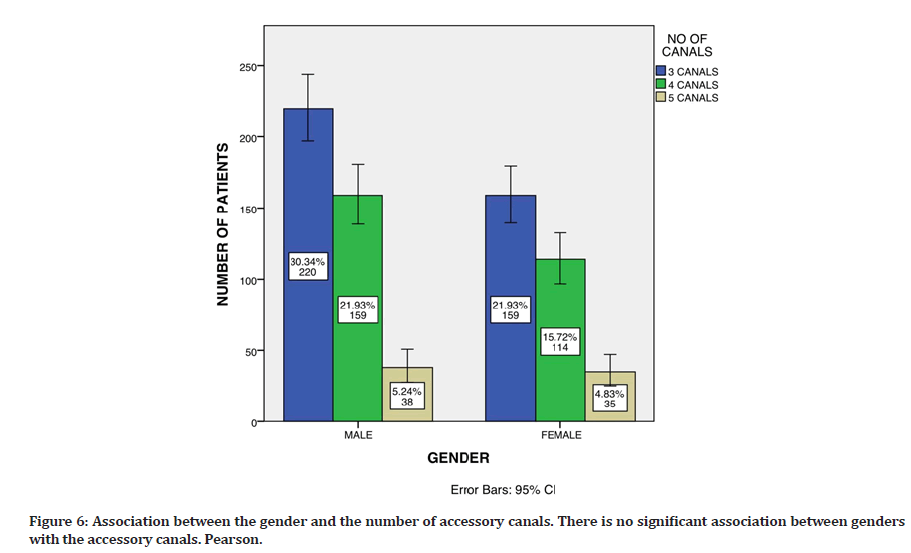
Figure 6: Association between the gender and the number of accessory canals. There is no significant association between genders with the accessory canals. Pearson.
Discussion
Primary molars in few studies show variations based on gender predilection, whereas in some studies there was no significant difference between gender. Some studies found the prevalence of an additional root had male predominance; others reported no difference between the sexes or rather more in females [27].
The incidence of three-rooted primary mandibular molars did not differ with gender (p>0.05) in the present study, which agrees with results reported by Najim, et al. [28]. Two roots and three canal system is the most common type in thе represent study. The mesial root consists of two canals mesio buccal and mesial lingual whereas the distal root consists of one canal in most of the cases. Very rarely two canals are seen in the distal root. Addition of one root has the least prevalence [29] based on gender was reported in previous studies [30].
Conclusion
Males showed a more prevalence for variations in canal morphology compared to the female population. Knowledge and variations about the canal morphology is a mist for all clinicians to perform adequate canal cleaning and shaping.
References
- Winter GB. Abscess formation in connexion with deciduous molar teeth. Arch Oral Biol 1962; 7:373â??379.
- Moss SJ, Addelston H, Goldsmith ED. Histologic study of pulpal floor of deciduous molars. J Am Dent Assoc 1965; 70:372â??379.
- Ramakrishnan M. Working length determination in mandibular primary second molars-A retrospective study. Int J Dent Oral Sci 2019; 17:10â??14.
- Jain P, Balasubramanian S, Sundaramurthy J, et al. A cone beam computed tomography of the root canal morphology of maxillary anterior teeth in an institutional-based study in Chennai urban population: an in vitro study. J Int Soc Prevent Community Dent 2017; 7:S68.
- Mohd Ariffin S, Dalzell O, Hardiman R, et al. Root canal morphology of primary maxillary second molars: A micro-computed tomography analysis. Eur Arch Paediatr Dent 2020; 21:519-525.
- Ramakrishnan M, Niveditha MS, Gurunathan D. A short review on the root canal configuration of primary maxillary molars. Bioinformation 2020; 16:1033.
- Kumar VD. A scanning electron microscope study of prevalence of accessory canals on the pulpal floor of deciduous molars. J Indian Soc Pedodont Prev Dent 2009; 27:85.
- http://isrctn.com/
- Ringelstein D, Seow WK. The prevalence of furcation foramina in primary molars. Pediatr Dent 1989; 11:198-202.
- Gutmann JL. Prevalence, location, and patency of accessory canals in the furcation region of permanent molars. J Periodontol 1978; 49:21-26.
- Zuza EP, Toledo BEC, Hetem S, et al. Prevalence of different types of accessory canals in the furcation area of third molars. J Periodontol 2006; 77:1755â??1761.
- Avinash K, Malaippan S, Dooraiswamy JN. Methods of isolation and characterization of stem cells from different regions of oral cavity using markers: A systematic review. Int J Stem Cells 2017; 10:12â??20.
- Pratha AA, Thenmozhi MS. A study of occurrence and morphometric analysis on meningo orbital foramen. Res J Pharm Technol 2016; 9:880â??882.
- Nair M, Jeevanandan G, Vignesh R. Comparative evaluation of post-operative pain after pulpectomy with k-files, kedo-s files and mtwo files in deciduous molars-a randomized clinical trial. Braz Dent J Â 2018; 21:411-417.
- Kannan R, Thenmozhi MS. Morphometric study of styloid process and its clinical importance on eagle’s syndrome. Res J Pharm Technol 2016; 9:1137â??1139.
- Samuel AR, Thenmozhi MS. Study of impaired vision due to Amblyopia. J Pharm Res 2015; 8:912-914.
- Viswanath A, Ramamurthy J, Dinesh SPS, et al. Obstructive sleep apnea: Awakening the hidden truth. Niger J Clin Pract 2015; 18:1â??7.
- Dinesh SPS, Arun AV, Sundari KKS, et al. An indigenously designed apparatus for measuring orthodontic force. J Clin Diagn Res 2013; 7:2623â??2626.
- Varghese SS, Thomas H, Jayakumar ND, et al. Estimation of salivary tumor necrosis factor-alpha in chronic and aggressive periodontitis patients. Contemp Clin Dent 2015; 6:152â??156.
- Priyanka S, Kaarthikeyan G, Nadathur JD, et al. Detection of cytomegalovirus, Epstein-Barr virus, and torque teno virus in subgingival and atheromatous plaques of cardiac patients with chronic periodontitis. J Indian Soc Periodontol 2017; 21:456â??460.
- Panda S, Jayakumar ND, Sankari M, et al. Platelet rich fibrin and xenograft in treatment of intrabony defect. Contemp Clin Dent 2014; 5:550â??554.
- Wu F, Zhu J, Li G, et al. Biologically synthesized green gold nanoparticles from Siberian ginseng induce growth-inhibitory effect on melanoma cells (B16). Artif Cells Nanomed Biotechnol 2019; 47:3297â??3305.
- Dua K, Wadhwa R, Singhvi G, et al. The potential of siRNA based drug delivery in respiratory disorders: Recent advances and progress. Drug Dev Res 2019; 80:714â??730.
- Patil SB, Durairaj D, Suresh Kumar G, et al. Comparison of extended nasolabial flap versus buccal fat pad graft in the surgical management of oral submucous fibrosis: A prospective pilot study. J Maxillofac Oral Surg 2017; 16:312â??321.
- Uthrakumar R, Vesta C, Raj CJ, et al. Bulk crystal growth and characterization of non-linear optical bisthiourea zinc chloride single crystal by unidirectional growth method. Curr Appl Phys 2010; 10:548â??552.
- Jain SV, Vijayakumar Jain S, Muthusekhar MR, et al. Evaluation of three-dimensional changes in pharyngeal airway following isolated lefort one osteotomy for the correction of vertical maxillary excess: A prospective study. J Maxillofac Oral Surg 2019; 18:139â??146.
- Lim HC, Jeon SK, Cha JK, et al. Prevalence of cervical enamel projection and its impact on furcation involvement in mandibular molars: A cone-beam computed tomography study in koreans. Anat Rec 2016; 299:379â??384.
- Najim U, Slotte C, Norderyd O. Prevalence of furcation-involved molars in a Swedish adult population. A radiographic epidemiological study. Clin Exp Dent Res 2016; 2:104â??111.
- Hou GL, Hung CC, Tsai CC, et al. Topographic study of extracted molars with advanced furcation involvement: Furcation entrance dimension and molar type. Kaohsiung J Med Sci 2003; 19:68â??73.
- Northway WM, Wainright RL, Demirjian A. Effects of premature loss of deciduous molars. Angle Orthod 1984; 54:295-329.
Indexed at, Google Scholar, Cross Ref
Indexed at, Google Scholar, Cross Ref
Indexed at, Google Scholar, Cross Ref
Indexed at, Google Scholar, Cross Ref
Indexed at, Google Scholar, Cross Ref
Indexed at, Google Scholar, Cross Ref
Indexed at, Google Scholar, Cross Ref
Indexed at, Google Scholar, Cross Ref
Indexed at, Google Scholar, Cross Ref
Indexed at, Google Scholar, Cross Ref
Indexed at, Google Scholar, Cross Ref
Indexed at, Google Scholar, Cross Ref
Indexed at, Google Scholar, Cross Ref
Indexed at, Google Scholar, Cross Ref
Indexed at, Google Scholar, Cross Ref
Indexed at, Google Scholar, Cross Ref
Indexed at, Google Scholar, Cross Ref
Indexed at, Google Scholar, Cross Ref
Indexed at, Google Scholar, Cross Ref
Indexed at, Google Scholar, Cross Ref
Indexed at, Google Scholar, Cross Ref
Author Info
Cibikkarthik T and Mahesh Ramakrishnan*
Department of Pedodontics, Saveetha Dental College and Hospitals, Saveetha Institute of Medical and Technical Sciences, Saveetha University, Tamil Nadu, IndiaCitation: Cibikkarthik T, Mahesh Ramakrishnan, Prevalence of Accessory Canals in Primary Mandibular Molars-A Radiographic Analysis, J Res Med Dent Sci, 2023, 11 (1):49-54.
Received: 03-Dec-2022, Manuscript No. jrmds-22-78959; , Pre QC No. jrmds-22-78959(PQ); Editor assigned: 05-Dec-2022, Pre QC No. jrmds-22-78959(PQ); Reviewed: 20-Dec-2022, QC No. jrmds-22-78959(Q); Revised: 26-Dec-2022, Manuscript No. jrmds-22-78959(R); Published: 02-Jan-2023
