Research - (2022) Volume 10, Issue 3
Prevalence Study of Squamous Cell Carcinoma in the Tongue
Abulqassim Raheem Daham* and Layla Sabri Yas
*Correspondence: Abulqassim Raheem Daham, Department of Oral and maxillofacial pathology, Dental Teaching Hospital, College of Dentistry, University of Baghdad, Iraq, Email:
Abstract
SCC of the tongue accounts for 20-25% of all malignant oral carcinomas. It typically affects older men in their 50-90s. Patients typically present with painless indurated or ulcerated lesions in the lateral aspect of anterior two thirds of tongue. Known etiologies include tobacco use, alcohol, and exposure to UV light. Other etiologies include trauma, nutritional deficiency, syphilis, and/or poor oral hygiene. SCC of the tongue initially metastasizes to the ipsilateral subdigastric lymph nodes. Aim: The aim was to determine the frequency and distribution of oral squamous cell carcinoma (OTSCC) involving tongue among patients by studying biopsy specimens during the period (2008-2020) years. Results: Of total cases of the tongue lesion (1451), 999 cases of squamous cell carcinoma in tongue region, The patient were affected over a wide range of 9–98 years with mean age of (54.98)years .The most age group effected (448, 44.80 %) in (60+). (514, 51.45 %) of male gender and (485, 48.55%) of female gender. The ratio of male to female is (1.05:1). Lateral border of the tongue was most commonly involved (451, 45.1%), followed by base of tongue and dorsal surface of tongue. Histopathological Grading, grade II (47.9%) had the highest percentage of the total number of cases (999) while grade IV were reported with the lowest percentage (3.1%). Baghdad governorate preponderance for the other governorates (646). 2020 year is the lowest value (28) cases as compared to the other years.
Keywords
Squamous cell carcinoma, Epidemiological study, Tongue
Introduction
Oral cancer is the sixth most common cancer worldwide [1,2]. More than 90% of all oral cancers are squamous cell carcinoma (SCC) [3]. Tongue squamous cell carcinoma (SCC) is the most common type of malignant tumor of the oral cavity, and usually occurs following the fifth decade of life [4]. Most cases occurring on the lateral border of the tongue. SCC of the tongue dorsum is very rare, especially at the midline [5]. The reasons why the lateral border of the tongue is the predilection sites for SCC because the carcinogens in the oral cavity mixing with saliva have the tendency to pool at the bottom of the mouth and these site are covered by thin and non-keratinized mucosa. As a consequence, they provide less protection against the carcinogen [6]. Epidemiological data shows that carcinoma of the tongue constituted 0.8% of all cancers in men and .04% of all cancers in women in United States.
Carcinoma of the tongue clinically presents as ulceration, fumigation (an exophytic mass), and infiltration with varying degree of induration. Patients rarely present with dysphagia or difficulty in speech. There is often a leukoplakic (white patch) or erythroplakic (red patch) component to the lesion and a small size does not exclude invasion [7,8].
The prognosis is strongly correlated with the stage of the disease at diagnosis. Survival of patients with stage I disease exceeds 80% [9]. For patients with locally advanced disease at the time of diagnosis (i.e., stage III and IV), survival drops below 40% [10]. The development of metastases in lymph nodes reduces the survival of a patient with a small primary tumor by 50%. Most patients with head and neck cancers at the time of diagnosis are found to be stage III or IV [11].
SCC of the tongue is usually graded by the histopathologists on its degree of differentiation into well, moderate, poor and undifferentiated squamous cell carcinoma. This is of use for oral oncologists and surgeons because there has been a correlation between histopathology and prognosis. This finding has got a great bearing on prognosis and 5 year survival because the prognosis for poorly differentiated and undifferentiated tumours is poor as compared to well differentiated tumours [12] (Figures 1 and Figure 2).
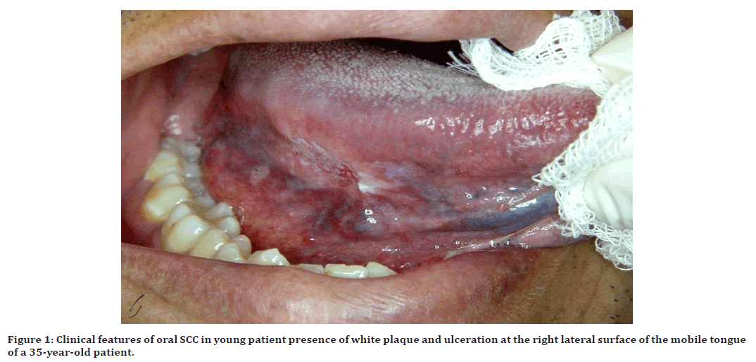
Figure 1. Clinical features of oral SCC in young patient presence of white plaque and ulceration at the right lateral surface of the mobile tongue of a 35-year-old patient.

Figure 2. A 73-year-old woman presented with a 1-year history of a persistent ulcer and white patch on the right margin of the tongue and soreness while eating.
Methodology
Study design
The related cases are collected from different hospitals of Iraq during the period 2008 to 2020 in the following confirmed centers:
✓ Department of oral and maxillofacial pathology, college of dentistry, university of Baghdad.
✓ Cancer registry center at Ministry of health/ Iraq.
✓ Major hospitals in Baghdad and governorates.
Results
Age and gender
514 cases (51.5%) of tongue SCC were observed in men and 485cases (48.5%) were seen in women, with a male: female ratio of (1.05:0.94). The mean age in male was years, (9–97) while in female it was 55.64 years (9–98). Overall mean age was 54.98 years (range: 9–98 years), it can be clearly observed that the maximum value (448) was reported in (60+ age group) while the minimum value (15) was reported in (<15 age group) shown in Tables 1 and Figure 3.
| Age group | Gender | Total | M:F | |
|---|---|---|---|---|
| Male | Female | Ratio | ||
| <15 | 9(0.9) | 6(0.6) | 15(1.5) | 1.5:1 |
| (15-30) | 14(1.4) | 8(0.8) | 22(2.2) | 1.75:1 |
| (30-45) | 57(5.7) | 53(5.3) | 110(11.0) | 1.07:1 |
| (45-59) | 208(20.8) | 196(19.6) | 404(40.4) | 1.06:1 |
| 60+ | 226(22.6) | 222(22.2) | 448(44.8) | 1.01:1 |
| Total | 514(51.5%) | 485(48.5%) | 999(100%) | 1.05:1 |
| Mean | 54.53 | 55.64 | 54.98 | |
| SD | 16.63119 | 15.933 | 16.294 | |
Table 1: Gender distribution of OSCC cases in relation to age group.
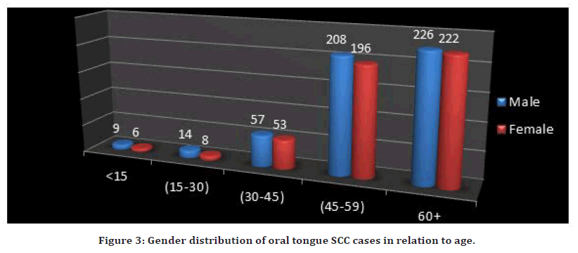
Figure 3. Gender distribution of oral tongue SCC cases in relation to age.
From Tables 1 and Figure 3, it can be observed that the maximum percentage (44.80 %) was found in age group (60+), while the minimum percentage (1.50 %) was found in age group (<15). 514 cases (51.5%) of tongue OSCC were observed in men and 485cases (48.5%) were seen in women, with a male: female ratio of (1.05:1). There was no significant relation between gender and age group (P=0.111575).
Prevalence and site anatomical
When anatomical sites were analyzed, the most commonly affected site was the lateral border the tongue (541 cases, 45.1%), followed base of the tongue (172 cases, 17.2%), dorsal surface of the tongue(69 cases, 6.9%),tip of tongue (46 cases, 4.6%), while 30 case was observed in the ventral surface of the tongue (30 case, 3.0%) ,It must be mentioned that (231 cases, 23.1%) cases were reported with tongue–NOS , it can be observed that the maximum percentage (45.1%) was found in Lateral border, while the minimum percentage (3.0%) was found in Ventral surface (Table 2 and Figure 4).
| ICD Code | N | % | ||
|---|---|---|---|---|
| Body of tongue | base | C01.9 | 172 | 17.2 |
| Dorsal surface | C02.0 | 69 | 6.9 | |
| Lateral border | C02.1 | 451 | 45.1 | |
| Ventral surface | C02.2 | 30 | 3 | |
| Tip | C02.1 | 46 | 4.6 | |
| tongue –NOS | C02.9 | 231 | 23.1 | |
| Total | 999 | 100 | ||
Table 2: Anatomical Site distribution for the tongue SCC cases.
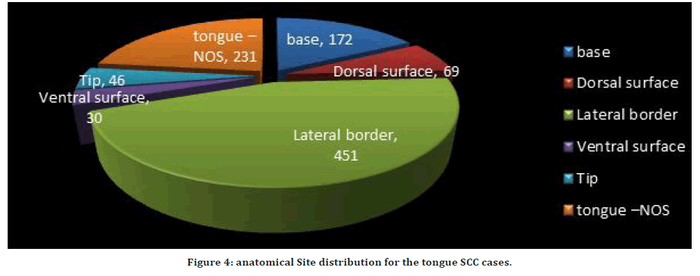
Figure 4. anatomical Site distribution for the tongue SCC cases.
The relative frequency of diagnostic categories of squamous cell carcinoma by annual patients
The data collection were gathered (999) cases, as squamous cell carcinoma that were recorded in (13) years in Iraqi governorates during the period 2008-2020: 2008 (53), 2009 (66), 2010 (78), 2011 (59), 2012 (84), 2013 (77), 2014 (108), 2015 (111), 2016 (104), 2017 (100), 2018 (99), 2019 (32), 2020 (28), as shown in Table 3. From Table 3, it can be clearly observed that the maximum value (111) was reported in 2015 year while the minimum value (28) was reported in 2020 year (Table 3 and Figure 5).
| Year | N | (%) | |
|---|---|---|---|
| 1 | 2008 | 53 | 5.3 |
| 2 | 2009 | 66 | 6.6 |
| 3 | 2010 | 78 | 7.8 |
| 4 | 2011 | 59 | 5.9 |
| 5 | 2012 | 84 | 8.4 |
| 6 | 2013 | 77 | 7.7 |
| 7 | 2014 | 108 | 10.8 |
| 8 | 2015 | 111 | 11.1 |
| 9 | 2016 | 104 | 10.4 |
| 10 | 2017 | 100 | 10 |
| 11 | 2018 | 99 | 9.9 |
| 12 | 2019 | 32 | 3.2 |
| 13 | 2020 | 28 | 2.8 |
| Total | 999 | 100 | |
Table 3: The annual number of new cases of OTSCC from 2008 to 2020.
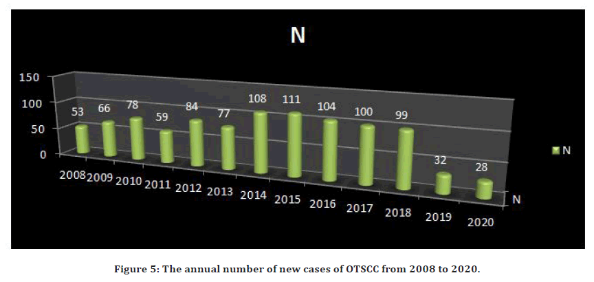
Figure 5. The annual number of new cases of OTSCC from 2008 to 2020.
Histopathological grading
The histopathological diagnosis of the OSCC indicated that, grade II represented by (47.9%) had the highest percentage of the total number of cases (999) then grade I (30.3%), grade III (4.7%) and the cases with grade IV were reported with the lowest percentage (3.1%). It must be mentioned that (139 , 13.9%) cases were reported with unstated (missing) differentiation, as in table 4, Moderately differentiated OTSCC was the most common (47.9%) among all histopathological grades while Undifferentiated, anaplastic(3.1%) was last percentage (Table 4 and Figure 6).
| WHO Grading | N | % | Histopathological Grading | |
|---|---|---|---|---|
| 1 | Grade I | 303 | 30.3 | Well differentiated, differentiated, NOS |
| 2 | Grade II | 479 | 47.9 | Moderately differentiated, Moderately well differentiated, intermediate differentiated |
| 3 | Grade III | 47 | 4.7 | Poorly differentiated |
| 4 | Grade IV | 31 | 3.1 | Undifferentiated , anaplastic |
| 9 | Not stated (missing) | 139 | 13.9 | Grade not determined, not stated or not applicable |
| Total | 999 | 100 |
Table 4: Distribution of histopathological grading for oral tongue SCC.
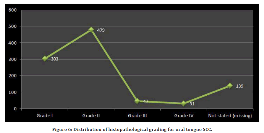
Figure 6. Distribution of histopathological grading for oral tongue SCC.
Iraq governorates
The data collection was collected (999) cases, as squamous cell carcinoma that were recorded in (10) Iraqi governorates: Baghdad (646), Basrah (69), Babil (56), Thiqar (50), Al-Najaf (40), Maysan (36), Al-Diwanyia (34), Karbala (28), Wasit (26) and Al-Muthanna (14), as shown in Table 5. From Table 5, it can be showed that the highest percentage (64.7%) was reported in Baghdad governorate while the lowest percentage (1.4%) was reported in Al-Muthanna governorate. The distribution of squamous cell carcinoma that it gathered for 10 governorates in Iraq from 2008 to 2020 is shown in Figure 7.
| No. | Governorate | Cases | Percentage (%) |
|---|---|---|---|
| 1. | Baghdad | 646 | 64.7 |
| 2. | Basrah | 69 | 6.9 |
| 3. | Babil | 56 | 5.6 |
| 4. | Thi qar | 50 | 5 |
| 5. | Al-Najaf | 40 | 4 |
| 6. | Maysan | 36 | 3.6 |
| 7. | Al-Diwanyia | 34 | 3.4 |
| 8. | Karbala | 28 | 2.8 |
| 9. | Wasit | 26 | 2.6 |
| 10. | Al-Muthanna | 14 | 1.4 |
| Total | 999 | 100 |
Table 5: Iraqi governorates distribution for oral tongue SCC cases.
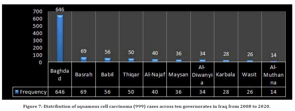
Figure 7. Distribution of squamous cell carcinoma (999) cases across ten governorates in Iraq from 2008 to 2020.
Methods
Data collection tools
The data collection for this study were collected from the filing procedure and archives of previously mentioned above during the period 2008 to 2020 using data collection sheet (case sheet) organized and generated for the purpose of this study. The processed case sheet collected data comprising:
Demographic characteristics of the cases (Patients numbers, age, gender and private information).
Tumor associated information:
Anatomical site and clinical behavior.
Reviewing of the patient recorders in different hospitals that are available.
Data were converted into a computerized database format. The database was checked for errors utilizing range and logical data cleaning techniques, and inconsistencies were remedied. An expert statistical advice was sought for statistical analyses were done using statistical package for social sciences (SPSS) version 21-computer software in association with Microsoft Excel 2013. Frequency distribution and percentages for selected variables describing the recorded cases with squamous cell carcinoma were done.
Male to female ratio has been used as indices for magnitude of gender difference. Thus, the Relative Risk (RR or risk ratio) is another index of effect size similar to its interpretation to standardized ratio.
RR = IR of risk group / IR of reference category.
Statistical analysis
Statistical analyses were performed using SPSS statistical package for Social Sciences (version 20.0 for windows, SPSS, Chicago, IL, USA). Qualitative data are represented as count and percentage. Chi-square test was used to test the relation of qualitative data. Quantitative data are represented as mean and standard deviation. P value of <0.05 was considered statistically significant. P>0.05 is the probability that the null hypothesis is true. A statistically significant test result (P ≤ 0.05) means that the test hypothesis is false or should be rejected. A P value greater than 0.05 means that no effect was observed.
Discussion
Age
Among the collected 999 OTSCC cases, the most affected age group was (60+yrs) (44.80%) which showed the highest percentage during the whole period from 2008-2020 in comparison to other age groups. This is in agreement with many of Iraqi studies [13,14] (56.6%); [15] (54.3%) ; Talabani et al. [16] (61.6%) and compatible with Khudier, 2012 and Museedi and Younis, 2014 . Also agreed with many international studies Shenoi et.al. [17] (42.7%); (88.3%) and consistent with [18,19].
Gender
OTSCC affects more frequently men than women. In a study conducted in UAE the M:F ratio was found to be (4:1.3) [19]. In USA, men were 2–4 times those among women [20]. Warnakulasuriya [21] in 2009 found that M:F is 1.5:1 which is most probably because more men than women face the high-risk habits. In Iran, male to female ratio is (2:1) [22]. Also in Canada the overall ratio of males to females with OC is (2:1) [23] and in India the male to female ratio is (2.2:1) [24,] and (4.18:1) according to [25]. In Germany, M: F (3 :1) by [26]. Our result were compatible with the previously mentioned international studies in that the overall male to female ratio was (1.4 : 1). Also this is in agreement with many Iraqi studies in reporting that male was more than female with different ratios (1.35:1, 1.2:1, 2:1, 1.2:1, 1.5:1,1.2:1, 1.3:1) according to [13,16,25,27,28] respectively. In Nigeria found a male to female ratio (3:4) that may be attributed to the increasing exposure of females to carcinogens such as tobacco and alcohol [29]. The difference in OC between men and women can be attributed to an increase in exposure of men to exogenous carcinogens. The variations in the contributions of smoking and alcohol were the possible causes of differences in OC between both genders.
Anatomical site
Out of the total cases (999) with OTSCC of the tongue our result showed that the lateral border of tongue was reported with the highest percentage (45.1%). These results were in consistence with (Hassan , 2008) who reported that the lateral border of tongue was the most common affected subsite (16.1%) and disagreed with Al-Reyahi et al. [13] tongue –NOS (64.9%).
Histopathological grading
Our results reported that the grade II represented by (47.9%) had the highest percentage out of the total number of cases (999). These results were in agreement with Hassan, 2008 and consistent with Hernández-Guerrero [20,29] (68.8%). This disagreed with [13,19,30-34] who reported that grade I was the most common grade. Also disagreed with [35,36] who reported that grade III was the most common grade. In addition to that, the determination of the grade of OTSCC is a subjective point and depend on the opinion of the histopathologist in his diagnosis and interpretation of the slide and this is may be an additional underlying cause for the high percentage of grade II .
It must be mentioned that (13.9%) cases were reported with unstated (missing) differentiation. Lack of information concerning the grade of OTSCC in a considerable number of cases in this study can be attributed to bad archiving of patients files and under registration in the providing centers of data. On the other side, some histopathological reports did not mention the differentiation. Also the histopathological slides for diagnosis were missed and not available specially the slides of the older years. The prognosis and treatment depends on the histological grade of the lesion as well as the clinical staging (TNM classification), and the age of the patient. Treatment typically involves surgical resection with radiation therapy. 5-year survival rate is about 20-30 (“Squamous cell carcinoma of tongue | Pathology Residency and Fellowship Program | Brown University”, n.d.).
Geographical distribution
The geographical distribution of the reported cases showed that the highest value of frequency distribution and percentage of cases was reported in Baghdad governorate (64.7%) during the study period (2008-2020). This is in agreement with all Iraqi researches that studied the geographical distribution of OC [13].
The high number of population can explain the underlying cause behind the highest percentages of OTSCC in Baghdad among other Iraqi governorates. Also the presence of many centers and hospitals, the location of the institute and hospital of radiotherapy and nuclear medicine center in Baghdad which made the referral cases reach the institute easier.
The lowest percentage was present in Al Muthanna and Karbala that is in agreement with [37]. This can be explained by the low number of population in these governorates in comparison to the high number of population in Baghdad .Also may be due to under registration of cases or loss of patient’s files and reports.
Conclusion
✓ The maximum percentage (44.80 %) was found in age group (60+), while the minimum percentage (1.50 %) was found in age group (<15).
✓ The males were more affected by OTSCC than females with overall male to female ratio (1.05:0.94).
✓ The most commonly affected site was the lateral border the tongue (541 cases, 45.1%) whereas the least affected site was the ventral surface of the tongue (30 cases, 3.0%).
✓ Moderately differentiated OTSCC was the most common (47.9%) among all histopathological grades followed by well differentiated OSCC (30.3%).
✓ The highest percentage (64.7%) was reported in Baghdad governorate while the lowest percentage (1.4%) was reported in Al-Muthanna governorate.
✓ The maximum cases (111) were reported in 2015 year while the minimum cases (28) were reported in 2020 year.
References
- https://www.brown.edu/academics/biomed/departments/pathology/residency/digital-pathology-library/head-and-neck/oral-cavity-tonsils-and-neck/squamous-cell-carcinoma-tongue
- Shah JP, Gil Z. Current concepts in management of oral cancer–surgery. Oral Oncol 2009; 45:394-401.
- Attar E, Dey S, Hablas A, et al. Head and neck cancer in a developing country: A population-based perspective across 8 years. Oral Oncol 2010; 46:591-596.
- Son YH, Kapp DS. Oral cavity and oropharyngeal cancer in a younger population. Review of literature and experience at Yale. Cancer 1985; 55:441-444.
- Goldenberg D, Ardekian L, Rachmiel A, et al. Carcinoma of the dorsum of the tongue. Head Neck 2000; 22(2):190-194.
- Johnson NW. Orofacial neoplasms: Global epidemiology, risk factors and recommendations for research. Int Dent J 1991; 41:365-75.
- Conley J, Sachs ME, Parke RB. The new tongue. Otolaryngol Head Neck Surg 1982; 90:58-68.
- Prince S, Bailey BM. Squamous carcinoma of the tongue. Br J Oral Maxillofac Surg 1999; 37:164-174.
- Siegel R, Ma J, Zou Z, et al. Cancer statistics, 2014. CA: Cancer J Clin 2014; 64:9-29.
- Harrison LB, RB Sessions, Hong WK. Head and neck cancer: A multidisciplinary approach. 3rd Edn. Philadelphia, PA.: Lippincott, William and Wilkins 2009.
- Wallner PE, Hanks GE, Kramer S, et al. Patterns of care study. Analysis of outcome survey data-anterior two-thirds of tongue and floor of mouth. Am J Clin Oncol 1986; 9:50-57.
- Khan M, Khitab U. Histopathological gradation of oral squamous cell carcinoma in niswar (snuff) dippers. Pakistan Oral Dent 2005; 25:173-176.
- Al-Reyahi AB. Retrospective analysis of malignant oral lesions for 1534 patients in Iraq during the period (1991–2000). A master thesis. Department of Oral diagnosis, College of Dentistry, University of Baghdad. 2004.
- Raja H AJ, Ghufran Adil H. Epidemiological of oral squamous cell carcinoma and salivary gland tumors using international classification of diseases for oncology (a cross-sectional study in Baghdad during the period 1999-2006).
- http://www.wales.nhs.uk/sites3/page.cfm?orgid=242&pid=59080
- Talabani NG, Ahmed KM, Faraj FH. Oral cancer in sulaimani: A clinicopathological study. J Zankoy Sulaimani 2008; 13:1-8.
- Shenoi R, Devrukhkar V, Sharma BK, et al. Demographic and clinical profile of oral squamous cell carcinoma patients: A retrospective study. Indian J Cancer 2012; 49:21.
- http://www.wales.nhs.uk/sites3/page.cfm?orgid=242&pid=59080
- Anis R, Gaballah K. Oral cancer in the UAE: A multicenter, retrospective study. Libyan J Med 2013; 8.
- Brown LM, Check DP, Devesa SS. Oral cavity and pharynx cancer incidence trends by subsite in the United States: Changing gender patterns. J Oncol 2012; 2012.
- Warnakulasuriya S. Global epidemiology of oral and oropharyngeal cancer. Oral Oncol 2009; 45:309-316.
- Mousavi SM, Gouya MM, Ramazani R, et al. Cancer incidence and mortality in Iran. Annals Oncol 2009; 20:556-63.
- Laronde DM, Hislop TG, Elwood JM, et al. Oral Cancer. Just the Facts. Clin Pract 2008; 74:269-272.
- Sharma P, Saxena S, Aggarwal P. Trends in the epidemiology of oral squamous cell carcinoma in Western UP: An institutional study. Indian J Dent Res 2010; 21:316.
- Khudier HH. Malignant oral lesions in Sulaimania governorate/a (5 years) retrospective study. Diyala J Med 2012; 2:13-20.
- Udeabor SE, Rana M, Wegener G, et al. Squamous cell carcinoma of the oral cavity and the oropharynx in patients less than 40 years of age: a 20-year analysis. Head Neck Oncol 2012; 4:1-7.
- Al-Niaimi AI. Oral malignant lesions in a sample of patients in the north of Iraq (Retrospective study). Al-Rafidain Dent J 2006; 6:176-80.
- Museedi OS, Younis WH. Oral cancer trends in Iraq from 2000 to 2008. Saudi J Dent Res 2014; 5:41-7.
- Lawal AO, Kolude B, Adeyemi BF. Oral cancer: The Nigerian experience. Int J Med Med Sci 2013; 5:178-83.
- Hernández-Guerrero JC, Jacinto-Alemán LF, Jiménez-Farfán MD, et al. Prevalence trends of oral squamous cell carcinoma. Mexico city’s general hospital experience. Med Oral Patol 2013; 18:e306.
- Al-Rawi NH, Talabani NG. Squamous cell carcinoma of the oral cavity: A case series analysis of clinical presentation and histological grading of 1,425 cases from Iraq. Clin Oral Investigations 2008; 12:15-18.
- Souza LR, Fonseca-Fonseca T, Oliveira-Santos CC, et al. Lip squamous cell carcinoma in a Brazilian population: Epidemiological study and clinicopathological associations. Med Oral Patol Oral Cir Bucal 2011; 16:757-762.
- Razmpa E, Memari F, Naghibzadeh B. Epidemiologic and clinicopathologic characteristics of tongue cancer in Iranian patients. Acta Medica Iranica 2011; 44-48.
- Halboub E, Al-Mohaya M, Abdulhuq M, Al-Mandili A, et al. Oral squamous cell carcinoma among Yemenis: Onset in young age and presentation at advanced stage. J Clin Exp Dent 2012; 4:e221.
- Effiom OA, Adeyemo WL, Omitola OG, et al. Oral squamous cell carcinoma: A clinicopathologic review of 233 cases in Lagos, Nigeria. J Oral Maxillofac Surg 2008; 66:1595-1599.
- Abdulai AE, Nuamah IK. Incidence of squamous cell carcinoma of the oral cavity and oropharynx in Ghanaians:-A retrospective study of histopathological charts in a teaching hospital. World J Surg Med Radi Oncol 2013; 3:75-83.
- Al-Kawaz AB. Oral squamous cell carcinoma in Iraq: Clinical analysis. MDJ 2010; 7.
Indexed at,Google Scholar, Cross Ref
Indexed at,Google Scholar, Cross Ref
Indexed at,Google Scholar, Cross Ref
Indexed at,Google Scholar, Cross Ref
Indexed at,Google Scholar, Cross Ref
Indexed at,Google Scholar, Cross Ref
Indexed at,Google Scholar, Cross Ref
Indexed at,Google Scholar, Cross Ref
Indexed at,Google Scholar, Cross Ref
Indexed at,Google Scholar, Cross Ref
Indexed at,Google Scholar, Cross Ref
Indexed at,Google Scholar, Cross Ref
Indexed at,Google Scholar, Cross Ref
Indexed at,Google Scholar, Cross Ref
Indexed at,Google Scholar, Cross Ref
Indexed at,Google Scholar, Cross Ref
Indexed at,Google Scholar, Cross Ref
Indexed at,Google Scholar, Cross Ref
Indexed at,Google Scholar, Cross Ref
Indexed at,Google Scholar, Cross Ref
Indexed at,Google Scholar, Cross Ref
Indexed at,Google Scholar, Cross Ref
Author Info
Abulqassim Raheem Daham* and Layla Sabri Yas
Department of Oral and maxillofacial pathology, Dental Teaching Hospital, College of Dentistry, University of Baghdad, IraqReceived: 04-Mar-2022, Manuscript No. JRMDS-22-51576; , Pre QC No. JRMDS-22-51576 (PQ); Editor assigned: 07-Mar-2022, Pre QC No. JRMDS-22-51576 (PQ); Reviewed: 21-Mar-2022, QC No. JRMDS-22-51576; Revised: 25-Mar-2022, Manuscript No. JRMDS-22-51576 (R); Published: 31-Mar-2022
