Research - (2022) Volume 10, Issue 8
Radial and Ulnar Artery Dominance among Sudanese Population-Colour Doppler Ultrasound Study
Khalid M Taha1,2,3, Juman M Almasaad4,5, Abubaker Y Elamin6, Mohammed H Karrar Alsharif7*, Nagi M. Bakhit8 and Mamoun A Alfaki7
*Correspondence: Mohammed H Karrar Alsharif, Department of Basic Medical Science, College of Medicine, Prince Sattam Bin Abdulaziz University, Saudi Arabia, Email:
Abstract
Background: This study aims to describe the appearance of radial and ulnar arteries diameter by mean values and determine the possible dominance of arterial supply in radial or ulnar arteries. Knowledge of the different diameters of forearm vessels is one of the most essential factors in coronary bypass surgery and radial artery conduit. Methods: The study was conducted on 28 individuals (14 male, 14) females with a mean age of 30, 5 years ± 5, 5 selected systematically by systematic sample technique. Patient prepared for arterial evaluation using color Doppler Ultrasound after 10 minutes rest on a couch. There was no hazard affecting the patients when using colour Doppler ultrasound. All patients have informed consent for ethical clearance. Results: The results showed that the mean diameter of right and left radial arteries were (2.5 ±.39) SD was (2.4 ±0.41) SD mm, (2.1 ± .29) SD mm, respectively, and right-left ulnar arteries diameter was (2.4 ± .41) SD mm, (2.2 ± .43) mm respectively. The dominant arterial supply was the right radial artery. Conclusions: There was no wide variation in mean diameter between radial and ulnar arteries in both hands because most samples were educated using their right hand rather than left. Subsequently, the dominant arterial supply of the hand was the right radial artery.
Keywords
Radial artery, Ulnar artery, Dominance, Diameter, Color doppler ultrasound
Introduction
The radial artery passes distally medial to the biceps tendon, across the supinator, over the tendon of insertion of the pronator teres, the radial origin of flexor digitorum superficial is, the origin of the flexor policies longus, the insertion of pronator quadratus, and the lower end of the radius. It disappears beneath the tendons of abductor policies longus and extensor policies brevis to cross the anatomical snuff box. The upper part of the forearm is overlapped anteriorly by the brachioradialis. The ulnar artery enters the hand anterior to the flexor retinaculum between the pisiform and the hook of the hamate via the ulnar canal (Guyon canal). The ulnar artery lies lateral to the ulnar nerve. The artery divides into two terminal branches, the superficial palmar arch and the deep palmar branch. The superficial palmar arch, the ulnar artery's main termination, gives rise to three common palmar digital arteries that anastomose with the palmar metacarpal arteries from the deep palmar arch.
The radial artery (RA) has emerged as an important arterial graft for coronary bypass surgery. With improving five-year patency rates [1] and increasing uptake, great attention has been focused on the optimal conduit harvesting technique after the internal thoracic artery [2]. Carpentier first introduced this method in 1973 but soon abandoned it due to the high rate of failure [3], .and this failure, due to the technique of preparations of the RA, had been responsible for graft failure [3]. There are different reasons to use the radial artery as a conduit: its caliber is similar to that of major coronary arteries (LAD); it has adequate thickness and resistance to the arterial wall and sufficient length to allow complete myocardial revascularization [4]. In the past, in preoperative screening, Allen's test was used to assess the blood flow in hand to protect from hand ischemia [5]. However, none of these tests helps evaluate the diameter and morphological appearance of the vessels with advanced knowledge. Nowadays, Doppler ultrasound is introduced as a possible method because it is non-invasive and accurate. Moreover, it allows morphological as well as functional evaluation of the forearm circulation [6]. This study aims to describe the appearance of radial and ulnar artery diameter by mean values and determine the possible dominant arterial supply of the forearm in radial or ulnar arteries.
Justifications
A hand complication after radial artery conduit for coronary bypass surgery, patients may lose hand capability due to arterial dominance of the hand. To reduce these complications, this study aims to give a clue about the forearm arteries diameters and then possible dominant artery in the hand.
Materials and Methods
Study design
Across sectional study design was performed from March 2021 to August 2021 on 28 individuals who came to the radiology department in Saheroon Special Hospital.
The study area
Special Saheroon Hospital is one of the most popular hospitals in Khartoum; it comprises many divisions' surgery, pediatric, medicine, anesthesia, and radiology departments. The radiology department consists of the most sophisticated tools never seen in other hospitals, especially colour Doppler ultrasound.
Patient preparation
A consecutive 28 Patients (14 males / 14 females) suitable for radial artery conduit were prepared according to the study protocol. The patient lay down on a couch for 5- 10 minutes of rest with a slightly flexed wrist. The hyperflexion of the wrist should be prevented; the significance of a slightly flexed wrist maneuver, the blood flow will be constant; no change occurs in vessels diameters.
All examinations were performed using an Aloka Prosdun 5SD- 35Sky ultrasound scanner (General Electric Medical Systems, Milwaukee, WI, USA) with a 9–14 MHz multi-frequency matrix linear transducer.
The right and left wrist was examined by resting the patient on the couch for ten minutes to reduce patient tension, and then a few Mg of ultrasound gill was put on the front of the wrist joint of both hands; a linear transducer prop was used to examine the forearm arteries.
The measurement site was from two fingerbreadths above the distal wrist crease; this procedure was repeated on both hands, then the mean of values was tacked as a final result.
All right-handed patients were included in this study; age, gender, and patient demographic data were considered. Patients with hand deformities or amputated upper limbs were excluded from this study.
Sample size
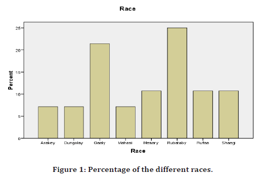
The sample size consists of 28 patients presenting in the radiology department at Special Saheroon Hospital. Most of the sample size shown below was Rubataby (Sudanese ethnic group) 25%, followed by Gaaly (Sudanese ethnic group) 22%.
Sample technique
The technique for sampling was systematic probability sampling.
Analysis
All data were collected in a design data collecting sheet; different statistical analyses were considered using SPSS INC 27. In addition, mean values were considered for arteries diameters.
Ethical considerations
After a full explanation by a researcher, all patients had informed consent that there was no harm obtained from US examinations; another paper request was performed to Special Saheroon Hospital's head manager to conduct such a study.
Results
In this study, consecutive 28 (14 males/14 females) persons suitable for radial artery conduit were presented in the radiology department in Special Saheroon Hospital; the result of the demographic data was tabulated in Table 1. Table 2 Shows the forearm arteries diameter shown in mm, their mean values, and SD, with significant values in the radial artery in both hands.
| Total number of Patients | 28 patient |
|---|---|
| Male\female | 14 male (50%)-14 female (50%) |
| Mean Age | 30.5 ± 5.5 SD |
| Smokers | 14.30% |
| Non Smokers | 85.70% |
| High Blood Pressure: Hypertensive | 14.30% |
| Non Hypertensive | 85.70% |
| Diabetic | 10.70% |
| Non-Diabetic | 89.30% |
| Write Handed Uses: Right-Handed | 82.1 |
| Left Handed | 17.9 |
Table 1: Patient demographic data.
| Arteries Diameter | n | Mean | SD |
|---|---|---|---|
| Right Radial Artery Diameter ~mm | 28 | 2.5 mm | 0.39 mm |
| Left Radial Artery Diameter~ mm | 28 | 2.4 mm | 0.41 mm |
| Right Ulnar Artery Diameter ~mm | 28 | 2.2 mm | 0.43 mm |
| Left Ulnar Artery ~Diameter mm | 28 | 2.1 mm | 0.29 mm |
Table 2: The mean diameter of both radial and ulnar arteries in both hands with their SD.
Radial and ulnar arteries diameter measurement
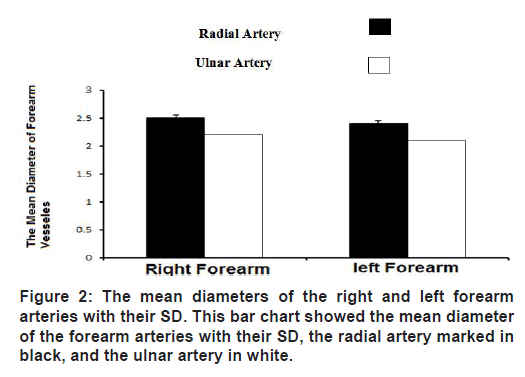
After the patient lay down on a bed for 5- 10 minutes of rest with a slightly flexed wrist, the hyper flexion of the wrist was prevented, and the radial and ulnar arteries diameter was measured on both sides. The results of the measurements revealed that the right radial artery diameter was 2.5 mm with SD 0.39 mm, while the left radial artery was 2.4 mm with SD 0 .41mm Figure 1 and Table 2. While the right ulnar artery diameter measurement was 2.2 mm with SD 0.43 mm, on the other hand, the left ulnar artery diameter was 2.1 mm with SD 29 mm and (Table 2).

Figure 1. Percentage of the different races.
Dominant side variation of the forearm arteries
In this study, most of the sample was right-handed people (using the right hand). This study revealed that the dominant hand supply was mainly the right radial artery and secondly by the left radial artery, which is slightly larger on the right side of the forearm, as seen in (Figure 2).

Figure 2. The mean diameters of the right and left forearm arteries with their SD. This bar chart showed the mean diameter of the forearm arteries with their SD, the radial artery marked in black, and the ulnar artery in white.
Radial and ulnar arteries dominant variations
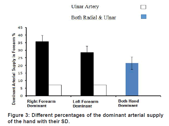
The radial artery (RA) has emerged as an important arterial graft for coronary bypass surgery. Dominant means the wide area in hand supplied by either radial or ulnar with considerable variations. This study revealed that the right radial artery was dominant (35.7%), followed by the left radial artery (28.57%). On the other hand, the right and left ulnar arteries' dominance was equal (7.14%), as seen in Figure 3.

Figure 3. Different percentages of the dominant arterial supply of the hand with their SD.
Gender dominant arterial supply variations
Gender means (male and female); this study considered gender an important variable depending on the sample type. The study revealed that the arterial supply of the hand was slightly more dominant in females by 0.1 mm than in males, as shown in Tables 3-6.
| Gender | Mean diameters in (mm) |
|---|---|
| Male | 2.5 |
| Female | 2.5 |
Table 3: The right radial artery diameter in gender.
| Gender | Mean diameters in (mm) |
|---|---|
| Male | 2.2 |
| Female | 2.2 |
Table 4: The right ulnar artery diameter in gender.
| Gender | Mean diameters in (mm) |
|---|---|
| Male | 2.3 |
| Female | 2.4 |
Table 5: The left radial artery diameter in gender.
| Gender | Mean diameters in (mm) |
|---|---|
| Male | 2.1 |
| Female | 2 |
Table 6: The left ulnar artery diameter in gender.
Discussion
This study revealed that the mean age was important when we measured the diameter of vessels, which tends to be smaller with advanced age [7]. Also, this study revealed that the mean diameter of the right and left radial was larger than the right and left ulnar arteries with their SD.
The radial artery passes downward and laterally beneath the brachioradialis muscle and rests on the forearm's deep muscles. In the middle third of its course, the radial nerve's superficial branch lies on its lateral side in the distal part of the forearm; the radial artery lies on the anterior surface of the radius and is covered only by skin and fascia. Here, the artery has a brachioradialis tendon on its lateral side and the tendon of flexor carpi radialis on its medial side (site for taking the radial pulse [8].
Upon entering the palm, the radial artery gives off the arteria radialis indices, which supply the lateral side of the index finger; the arteria princeps pollicis, which divides into two and supplies the lateral and medial sides of the thumb. The deep palmar arch is a direct continuation of the radial artery and supplies most of the deep tendons [9].
Moreover, the radial arteries are larger in diameter than the ulnar arteries. Subsequently, the right and left radial arteries are a little bit larger than the right and left ulnar arteries, which agreed with that published in the literature [10-12]; this finding was in disagreement with that published literature [13]; those authors said that the ulnar artery was larger than the radial artery at the level of the elbow, but in this study, was measured at the level of the wrist and the ulnar artery tend to be smaller because it gave many muscular branches at the wrist.
The diameter of radial and ulnar arteries varies in different populations, sometimes less than 2 mm, as a study conducted in Finland, and depends on the nature of people's work.
This study showed that the dominance of these arteries was more than 2 mm, and my sample toked from educated people, which was why they were less than 2mm [14]. This study showed no wide variations between the mean diameter of the radial and ulnar arteries. The dynamic studies included colour Doppler sonography done in 22 individuals (44 hands) and fivechannel plethysmography in 40 individuals (40 right hands). It revealed that the ulnar artery is dominant at the elbow. However, after giving its collateral branches, the radial artery becomes dominant in the distal forearm and constitutes the primary source of vascularization in hand. The ulnar artery is rarely dominant at the forearm level and is physiologically less important [15].
In this study, the dominant arterial supply was a right radial, followed by a left radial. The right and left radial arteries were dominant in this study due to the most data were the right-handed people. However, the P. value was more than 0.05; there was no significant correlation between the right forearm arteries diameter in conformity with that published literature [16]. In gender, the female artery diameter was slightly larger than male due to the type of work of females as the same as the male (educated people).
Conclusion
The right and left radial arteries' diameter was significantly larger than the right and left ulnar arteries, and the dominant arterial hand supply was the right radial artery. However, there was no significant correlation between the right dominance of the arteries and their diameters.
Acknowledgment
This publication was supported by the Deanship of Scientific Research at Prince Sattam bin Abdulaziz University, Alkharj, Saudi Arabia.
Authors Contributions
All authors contributed equally to this work and have read and agreed to the final manuscript.
Funding
This research received no external funding.
Conflict of Interest
The authors declare that they have no conflict of interest.
Other Disclosure
None.
References
- Acar C, Jebara VA, Portoghese M, et al. Revival of the radial artery for coronary artery bypasses grafting. Ann Thorac Surg 1992; 54:652-660.
- Blitz A, Osterday RM, Brodman RF. Harvesting the radial artery. Anna Cardiothorac Surg 2013; 2:533.
- https://link.springer.com/book/10.1007/3-540-30084-8
- Pola P, Serricchio M, Flore R, et al. Safe removal of the radial artery for myocardial revascularization: a Doppler study to prevent ischemic complications to the hand. J Thorac Cardiovasc Surg 1996; 112:737-744.
- Starnes S, Wolk W, Lampman M, et al. Noninvasive evaluation of hand circulation before radial artery harvest for coronary artery bypass grafting. J Thorac Cardiovasc Surg 1999; 117:261–266.
- Rodriguez E, Ormont ML, Lambert EH, et al. The role of preoperative radial artery ultrasound and digital plethysmography prior to coronary artery bypass grafting. Eur J Cardio-thoracic Surg 2001; 19:135-139.
- Huzjan R, Brkljačić B, Delić-Brkljačić D, et al. B-mode and color doppler ultrasound of the forearm arteries in the preoperative screening prior to coronary artery bypass grafting. Collegium Antropologicum 2004; 28:235-241.
- https://www.worldcat.org/title/grays-anatomy-the-anatomical-basis-of-clinical-practice/oclc/213447727
- https://www.textbooks.com/Clinical-Anatomy-by-Regions-8th-Edition/9780781764049/Richard-S-Snell.php
- Okuyan H, Hzal F. Angiographic evaluation of the radial artery diameter in patients who underwent coronary angiography or coronary intervention. J Invasive Cardiol 2013; 25.
- Alejandro Velasco MD, Chikako Ono MD, Kenneth Nugent MD, et al. Ultrasonic evaluation of the radial artery diameter in a local population from Texas. J Invasive Cardiol 2012; 24.
- Brzezinski M, Luisetti T, London MJ. Radial artery cannulation: A comprehensive review of recent anatomic and physiologic investigations. Anesth Analg 2009; 109:1763-1781.
- Doscher WI, Viswanathan BA, Stein TH, et al. Hemodynamic assessment of the circulation in 200 normal hands. Ann Surg 1983; 198:776.
- Kohonen M, Teerenhovi O, Terho T, et al. Non-harvestable radial artery. A bilateral problem. Interact Cardiovasc Thorac Surg 2008; 7:e800.
- Haerle M, Häfner HM, Dietz K, et al. Vascular dominance in the forearm. Plast Reconstr Surg 2003; 189- 192.
- Fuhrman TM, McSweeney E. Noninvasive evaluation of the collateral circulation to the hand. Acad Emerg Med 1995; 2:195-199.
Indexed at, Google Scholar, Cross Ref
Indexed at, Google Scholar, Cross Ref
Indexed at, Google Scholar, Cross Ref
Indexed at, Google Scholar, Cross Ref
Indexed at, Google Scholar, Cross Ref
Indexed at, Google Scholar, Cross Ref
Indexed at, Google Scholar, Cross Ref
Indexed at, Google Scholar, Cross Ref
Indexed at, Google Scholar, Cross Ref
Author Info
Khalid M Taha1,2,3, Juman M Almasaad4,5, Abubaker Y Elamin6, Mohammed H Karrar Alsharif7*, Nagi M. Bakhit8 and Mamoun A Alfaki7
1Department of Anatomy, Faculty of Medicine, University of Garden City, Sudan2Department of Anatomy, Faculty of Medicine, Omdurman Islamic University, Sudan
3Department of Anatomy, Faculty of Medicine El Deain, Sudan
4Department of Basic Medical Sciences, College of Medicine, King Saud Bin Abdul Aziz University for Health Sciences, National Guard Health Affairs, Jeddah, Saudi Arabia
5King Abdullah International Medical Research Centre (KAIMRC), King Abdulaziz Medical City, Jeddah, Saudi Arabia
6Department of Histology and Embryology, Faculty of Medicine, Ondokuz Mayis University, 55139 Atakum, Samsun, Turkey
7Department of Basic Medical Science, College of Medicine, Prince Sattam Bin Abdulaziz University, Al Kharj, 11942, Saudi Arabia
8Department of Anatomy, Arabian Gulf University, Manama, Bahrain
Received: 15-Jul-2022, Manuscript No. jrmds-22-71906; , Pre QC No. jrmds-22-71906(PQ); Editor assigned: 18-Jul-2022, Pre QC No. jrmds-22-71906(PQ); Reviewed: 26-Jul-2022, QC No. jrmds-22-71906; Revised: 29-Jul-2022, Manuscript No. jrmds-22-71906(R); Published: 05-Aug-2022
