Research - (2022) Volume 10, Issue 6
Retrospective Analysis on Treatment Modalities for Non-Vital Open Apex Cases
Baala Vignesh A and Pradeep S*
*Correspondence: Pradeep S, Department of Conservative Dentistry and Endodontics, Saveetha Dental College and Hospitals, Saveetha Institute of Medical and Technical Sciences (SIMATS), India, Email:
Abstract
Aim: To compare and evaluate the prevalence of non-vital open apex cases reported in Saveetha Dental College and compare it with the sealers used. Introduction: An immature permanent tooth having a blunderbuss canal and open apex can be an endodontic challenge because of difficulty in obtaining an apical seal, and existing thin radicular walls which are susceptible to fracture. One step apexification is defined as nonsurgical compaction of a biocompatible material into the apical end of the root canal to establish an apical stop that would enable the root canal to be immediately filled. Materials and Method: The study was conducted in Saveetha Dental College and Hospitals, with patients visiting for a period between September 2019 and march 2020. The data was collected by reviewing the case sheets. Patient age, gender, apexification procedure done, nolla stages, material used for apexification as per the records were collected. The obtained data was entered in Ms Excel spreadsheet and the tabulated data was subjected to statistical software IBM SPSS version 20.0. Descriptive inferential statistics were done. Chi Square test applied and the p value was set at p<0.05. Results: The study from the above results showed that out of 82 patients, 18 (15 MTA, 3 Biodentine) were between the age of 0-10yrs, 34 (25 MTA, 9 Biodentin) were between 10-20 and 26 (19 MTA and 7 Biodentin) were between the age 20-30 and 4 (3 MTA, 1 Biodentine) were between the age group 30-40. The most frequently used apexification material was MTA with a frequency of 62 patients to biodentine which was used in 20 patients. The study was statistically significant (P=0.042) with the highest frequency of open apexes seen with ages 10-20 and MTA was being used as an apical plug. Nolla’s stage 9 had maximum prevalence and was found to be statistically significant when compared to other stages (p<0.05). Maxillary central incisors had more apexification procedures as a result of trauma at the young age. Conclusion: The present study shows that the highest prevalence of open apexes were seen in patients between 10-20 years of age, the main reason was found to be trauma. MTA was used as a apexification material compared to biodentine. Hence further studies are required to obtain long term effects of various apexification procedures.
Keywords
Apexification, Biodentin, MTA, Nollas stages of development, Open Apex
Introduction
An immature permanent tooth having a blunderbuss canal and open apex can be an endodontic challenge because of difficulty in obtaining an apical seal, and existing thin radicular walls which are susceptible to fracture [1]. This was overcome by apexification, which was done with the help of MTA or bio dentin. One step apexification is defined as nonsurgical compaction of a biocompatible material into the apical end of the root canal to establish an apical stop that would enable the root canal to be immediately filled [2].
Several biocompatible materials such as dentin chips, bovine true ceramics, tricalcium phosphate, collagencalcium phosphate, bone growth factors, and oxidized cellulose have been used to create an apical matrix [3]. MTA has a remarkable capacity to induce hard-tissue formation and periradicular healing; however, the material may still get extruded into periapex in teeth with wide apices preventing its adequate compaction [4]. This material is autogenous, resorbable, promotes healing, has lesser allergic potential and provides a stable matrix against which an apical barrier is formed [5].
Biodentine is a new calcium silicate based cement of the same type as MTA. It exhibits physical and chemical properties similar to those described for certain Portland cement derivatives. Based on all its properties, Biodentin has been claimed to be a bioactive dentin substitute for the repair of root perforations, apexification and retrograde root filling by the manufacturers [6]. A modified powder composition, the addition of setting accelerators and softeners, and a new predosed capsule formulation for use in a mixing device, largely improved the physical properties of this material making it much more user-friendly with a shorter setting [7].
Thus this study compares the efficacy of MTA and biodentine used in the treatment of non-vital apex with apexification procedures, with the case reports of apexification procedures done in Saveetha Dental College.
Our team has extensive knowledge and research experience that has translate into high quality publications [8-26].
Materials and Methods
The study was conducted in Saveetha Dental College and Hospitals, with patients visiting for a period between September 2019 and march 2020. The data was collected by reviewing the case sheets. The study setting was approved by the Institutional ethics committee SDC/ SIHEC/2020/DIASDATA/0619-0320. Two examiners were involved in the study. Total of patients case sheets reviewed, Patient age, gender, apexification procedure done, nolla stages, material used for apexification as per the records were collected. Telephonic and photographic cross verification of data was done by two examiners. If there was no response from the patient, the particular data was excluded. The dependent variables and independent variables were set. The obtained data was entered in MS Excel spreadsheet and the tabulated data was subjected to statistical software IBM SPSS version 20.0. Descriptive inferential statistics were done. Chi Square test applied and the p value was set at p<0.05.
Results
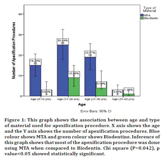
The study from the above results showed that out of 82 patients, 18 (15 MTA, 3 Biodentine) were between the age of 0-10yrs, 34 (25 MTA, 9 Biodentin) were between 10- 20 and 26 (19 MTA and 7 Biodentin) were between the age 20-30 and 4 (3 MTA, 1 Biodentine) were between the age group 30-40. The most frequently used apexification material was MTA with a frequency of 62 patients to biodentine which was used in 20 patients. The study was statistically significant chi square (P=0.042) with the highest frequency of open apexes seen with ages 10- 20 and MTA was being used as an apical plug. (Figure 1).

Figure 1. This graph shows the association between age and type of material used for apexification procedure. X axis shows the age and the Y axis shows the number of apexification procedures. Blue colour shows MTA and green colour shows Biodentine. Inference of this graph shows that most of the apexification procedure was done using MTA when compared to Biodentin. Chi square (P=0.042), p value<0.05 showed statistically significant.
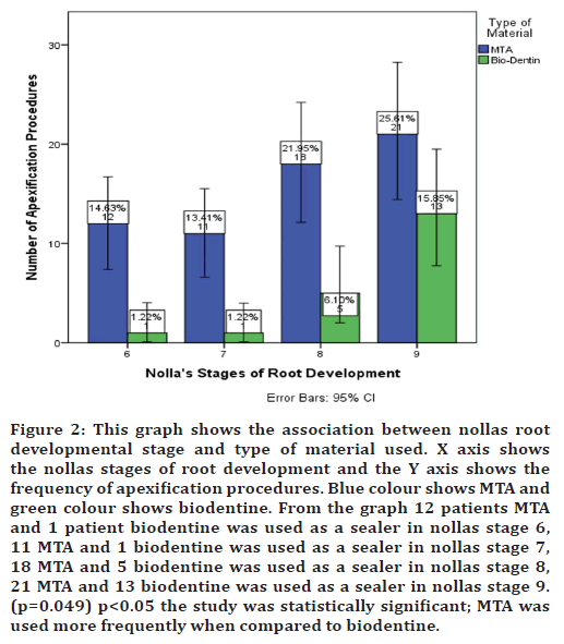
The association between nollas stage of root development and type of sealer material used showed that, 12 patients MTA and 1 patient biodentine was used as a sealer in nollas stage 6, 11 MTA and 1 biodentine was used as a sealer in nollas stage 7, 18 MTA and 5 biodentine was used as a sealer in nollas stage 8, 21 MTA and 13 biodentine was used as a sealer in nollas stage 9. Chi square (p=0.049) p<0.05, the study was statistically significant. Nollas stage 9 was seen with increased prevalence with MTA being used as an apical plug (Figure 2).

Figure 2. This graph shows the association between nollas root developmental stage and type of material used. X axis shows the nollas stages of root development and the Y axis shows the frequency of apexification procedures. Blue colour shows MTA and green colour shows biodentine. From the graph 12 patients MTA and 1 patient biodentine was used as a sealer in nollas stage 6, 11 MTA and 1 biodentine was used as a sealer in nollas stage 7, 18 MTA and 5 biodentine was used as a sealer in nollas stage 8, 21 MTA and 13 biodentine was used as a sealer in nollas stage 9. (p=0.049) p<0.05 the study was statistically significant; MTA was used more frequently when compared to biodentine.
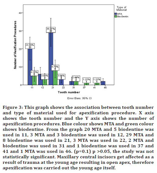
The association between open apex incidence and sealer material used showed that, 20 MTA and 5 biodentine was used in 11, 3 MTA and 3 biodentine was used in 12, 29 MTA and 8 biodentine was used in 21, 3 MTA was used in 22, 2 MTA and biodentine was used in 31 and 1 biodentine was used in 37 and 41 and 1 MTA was used in 46. Chi square (p=0.1) p >0.05 the study was not statistically significant. Most open apexes were seen in relation to 21 and MTA was used as an apical plug. (Figure 3).

Figure 3. This graph shows the association between tooth number and type of material used for apexification procedure. X axis shows the tooth number and the Y axis shows the number of apexification procedures. Blue colour shows MTA and green colour shows biodentine. From the graph 20 MTA and 5 biodentine was used in 11, 3 MTA and 3 biodentine was used in 12, 29 MTA and 8 biodentine was used in 21, 3 MTA was used in 22, 2 MTA and biodentine was used in 31 and 1 biodentine was used in 37 and 41 and 1 MTA was used in 46. (p=0.1) p >0.05, the study was not statistically significant. Maxillary central incisors get affected as a result of trauma at the young age resulting in open apex, therefore apexification was carried out the young age itself.
Discussion
The results of the present study showed that increased prevalence of open apexes were seen in patients between the age 10-20. The study by Kumar, et al showed that increased open apexes were seen in patients between 10-20 correlating with the present study [27]. The study by Sharma V, et al stated that increased open apexes are seen in patients of age group 5-10 which is contradictory to the present study [28]. The study by Cameriere, et al states that the 10 to 14-year-old age group was the group most commonly submitted to treatment (40.6%), as reported in previous studies and these procedures are not exclusive to young patients with immature roots [29]. Completely formed teeth can suffer alteration in the terminal portion of the root by pathological or iatrogenic factors and develop open apices.
The nollas stages of root development include [30]:
Stage 10: Apical end of root completed
Stage 9: Root almost complete; open apex
Stage 8: Two‐third of root completed
Stage 7: One‐third of root completed
Stage 6: Crown completed
Stage 5: Crown almost completed
Stage 4: Two‐third of crown completed
Stage 3: One‐third of crown completed
Stage 2: Initial calcification
Stage 1: Presence of crypt
stage 0: Absence of crown
A comparison of the nollas stages of root development and age showed that 48.72 % of nollas stage 9 was seen in patients of age 10-20. The study by Sinha, et al stated that nollas stage 9 of root development was seen with increased prevalence in patients of age groups 10-13 correlating with the present study [31].
The present study shows that MTA was the most commonly used sealing material used for apexification. The study by Betul Gomez, et al stated that MTA apical plug method is effective because of the less requirement of treatment time, appointments and radiographs, and better fracture resistance after the treatment of nonvital immature permanent teeth correlating with the study [32]. MTA consists of fine hydrophilic particles of tricalcium silicate, silicate oxide and tricalcium oxide. MTA has been suggested to create an apical plug at the root-end and helps to prevent the extrusion of the filling materials. MTA shows less leakage, better marginal adaptation with shortened setting time [33] It also shows hydrophilic characteristics, creating a favorable environment for the positive outcome for the treatment [34]. These autogenous materials are preferred over synthetic materials to avoid the possibility of treatment failure. MTA showed lesser inflammation, hyperemia, necrosis with more odontoblastic layer formation when compared with other cements [35]. Thus it can be used as an effective apical barrier in teeth with necrotic pulp and open apex findings [36]. But MTA remains subject to some concerns, such as its long setting time, poor handling characteristics, low resistance to compression, low flow capacity, limited resistance to washout before setting, possibility of staining of tooth structure, presence and release of arsenic, and high cost [37]. These disadvantages necessitate more ideal restorative materials, with adequate biological and mechanical properties. Biodentine is a newly developed calcium silicate-based material that has recently been introduced as a dentine substitute, whenever original dentine is damaged [38]. In contrast with MTA, the mechanical properties of Biodentine are similar to those of natural dentine. The compressive strength, elasticity modulus and microhardness are comparable with that of natural dentine [39]. The material is stable, less soluble, non-resorbable, hydrophilic, easy to prepare and place, needs much less time for setting, produces a tighter seal and has greater radiopacity [40]. Due to its improved material properties, Biodentine has a distinct advantage over its closest alternatives in treatment of teeth with open apex [41].
The future scope of study includes investigation of MTA to prove its long term efficacy. Better methods to incorporate proper seal in open apex cases is to be incorporated during treatment.
Conclusion
The present study shows that the highest prevalence of open apexes were seen in patients between 10-20 years of age, the main reason was found to be trauma. MTA was used as a apexification material compared to biodentine. Hence further studies are required to obtain long term effects of various apexification procedures.
References
- Alkhadra T, Preshing W, El-Bialy T. Prevalence of traumatic dental injuries in patients attending University of Alberta Emergency Clinic. Open Dent J 2016; 10:315-321.
- Petti S, Tarsitani G, Arcadi P, et al. The prevalence of anterior tooth trauma in children 6 to 11 years old. Minerva Stomatol 1996; 45:213-218.
- Afonso T, de Andrade Moura ML, Pega AL, et al. Effect of calcium hydroxide as intracanal medication on the apical sealing ability of mineral trioxide aggregate (MTA): an in vitro apexification model. J Health Sci Inst 2012; 30:104-112.
- Giuliani V, Baccetti T, Pace R, et al. The use of MTA in teeth with necrotic pulps and open apices. Dent Traumatol 2002; 18:217-221.
- Torabinejad M, Watson TF, Ford TP. Sealing ability of a mineral trioxide aggregate when used as a root end filling material. J Endod 1993; 19:591-595.
- Parirokh M, Torabinejad M, Dummer PM. Mineral trioxide aggregate and other bioactive endodontic cements: an updated overview–part I: Vital pulp therapy. Int Endod J 2018; 51:177-205.
- Pradelle-Plasse N, Tran XV, Colon P. Physico-chemical properties. Biocompatibility or cytotoxic effects of dental composites Oxford: Coxmoor. 2009;184-194.
- Muthukrishnan L. Imminent antimicrobial bioink deploying cellulose, alginate, EPS and synthetic polymers for 3D bioprinting of tissue constructs. Carbohydr Polym 2021; 260:117774.
- Kim DH, Lee S, Jung J, et al. Near-infrared autofluorescence-based parathyroid glands identification in the thyroidectomy or parathyroidectomy: A systematic review and meta-analysis. Langenbecks Arch Surg 2021; 28:1-9.
- Chakraborty T, Jamal RF, Battineni G, et al. A review of prolonged post-COVID-19 symptoms and their implications on dental management. Int J Environ Res Public Health. 2021 Jan;18(10):5131.
- Muthukrishnan L. Nanotechnology for cleaner leather production: A review. Environ Chem Lett 2021; 19:2527-2549.
- Teja KV, Ramesh S. Is a filled lateral canal–A sign of superiority?. J Dent Sci 2020; 15:562.
- Narendran K, MS N, Sarvanan A. Synthesis, Characterization, Free Radical Scavenging And Cytotoxic Activities Of Phenylvilangin, A Substituted Dimer Of Embelin. Indian J Pharm Sci 2020; 82:909-912.
- Reddy P, Krithikadatta J, Srinivasan V, et al. Dental caries profile and associated risk factors among adolescent school children in an urban South-Indian city. Oral Health Prev Dent 2020; 18:379-386.
- Sawant K, Pawar AM, Banga KS, et al. Dentinal microcracks after root canal instrumentation using instruments manufactured with different NiTi alloys and the SAF system: A systematic review. Appl Sci 2021; 11:4984.
- Bhavikatti SK, Karobari MI, Zainuddin SL, et al. Investigating the antioxidant and cytocompatibility of MimusopselengiLinn extract over human gingival fibroblast cells. Int J Environ Res Public Health. 2021; 18:7162.
- Karobari MI, Basheer SN, Sayed FR, et al. An In Vitro stereomicroscopic evaluation of bioactivity between Neo MTA Plus, Pro Root MTA, BIODENTINE & glass ionomer cement using dye penetration method. Materials 2021; 14:3159.
- Rohit Singh T, Ezhilarasan D. Ethanolic extract of Lagerstroemia Speciosa (L.) Pers., induces apoptosis and cell cycle arrest in HepG2 cells. Nutr Cancer 2020; 72:146-56.
- Ezhilarasan D. MicroRNA interplay between hepatic stellate cell quiescence and activation. Eur J Pharmacol 2020; 885:173507.
- Romera A, Peredpaya S, Shparyk Y, et al. Bevacizumab biosimilar BEVZ92 versus reference bevacizumab in combination with FOLFOX or FOLFIRI as first-line treatment for metastatic colorectal cancer: A multicentre, open-label, randomised controlled trial. Lancet Gastroenterol Hepatol 2018; 3:845-855.
- Vijayashree Priyadharsini J. In silico validation of the non‐antibiotic drugs acetaminophen and ibuprofen as antibacterial agents against red complex pathogens. J Periodontol 2019; 90:1441-1448.
- Priyadharsini JV, Girija AS, Paramasivam A. In silico analysis of virulence genes in an emerging dental pathogen A. baumannii and related species. Arch Oral Biol 2018; 94:93-98.
- Uma Maheswari TN, Nivedhitha MS, Ramani P. Expression profile of salivary micro RNA-21 and 31 in oral potentially malignant disorders. Braz Oral Res 2020; 34.
- Gudipaneni RK, Alam MK, Patil SR, et al. Measurement of the maximum occlusal bite force and its relation to the caries spectrum of first permanent molars in early permanent dentition. J Clin Pediatr Dent 2020; 44:423-428.
- Chaturvedula BB, Muthukrishnan A, Bhuvaraghan A, et al. Dens invaginatus: A review and orthodontic implications. Br Dent J 2021; 230:345-350.
- Kanniah P, Radhamani J, Chelliah P, et al. Green synthesis of multifaceted silver nanoparticles using the flower extract of Aerva lanata and evaluation of its biological and environmental applications. ChemistrySelect. 2020; 5:2322-2331.
- Kumar SM, Kumar T, Keshav V, et al. Open apex solutions: One-step apexification, salvaging necrosed teeth with open apex. Endodontology. 2019; 31:173.
- Sharma V, Sharma S, Dudeja P, et al. Endodontic management of nonvital permanent teeth having immature roots with one step apexification, using mineral trioxide aggregate apical plug and autogenous platelet-rich fibrin membrane as an internal matrix: Case series. Contemp Clin Dent 2016; 7:67-70.
- Cameriere R, Pacifici A, Pacifici L, et al. Age estimation in children by measurement of open apices in teeth with Bayesian calibration approach. Forensic Sci Int 2016; 258:50-54.
- Sachan K, Sharma VP, Tandon P. Reliability of Nolla's dental age assessment method for Lucknow population. J Pediatr Dent 2013; 1:8-13.
- Sinha S, Umapathy D, Shashikanth MC, et al. Dental age estimation by Demirjian's and Nolla's method: A comparative study among children attending a dental college in Lucknow (UP). J Indian Acad Oral Med Radiol 2014; 26:279.
- Güneş B, Aydinbelge HA. Mineral trioxide aggregate apical plug method for the treatment of nonvital immature permanent maxillary incisors: Three case reports. J Conserv Dent 2012; 15:73-76.
- Holden DT, Schwartz SA, Kirkpatrick TC, et al. Clinical outcomes of artificial root-end barriers with mineral trioxide aggregate in teeth with immature apices. J Endod 2008; 34:812-817.
- Çiçek E, Yılmaz N, Koçak MM, et al. Effect of mineral trioxide aggregate apical plug thickness on fracture resistance of immature teeth. J Endod 2017; 43:1697-1700.
- Oliveira TM, Sakai VT, Silva TC, et al. Mineral trioxide aggregate as an alternative treatment for intruded permanent teeth with root resorption and incomplete apex formation. Dent Traumatol 2008; 24:565-568.
- Schwartz RS, Mauger M, Clement DJ, et al. Mineral trioxide aggregate: A new material for endodontics. J Am Dent Assoc 1999; 130:967-975.
- Pinar Erdem A, Sepet E. Mineral trioxide aggregate for obturation of maxillary central incisors with necrotic pulp and open apices. Dent Traumatol 2008; 24:e38-41.
- Nayak G, Hasan MF. Biodentine-a novel dentinal substitute for single visit apexification. Restor Dent Endod 2014; 39:120-125.
- Tay FR, Pashley DH, Rueggeberg FA, et al. Calcium phosphate phase transformation produced by the interaction of the Portland cement component of white mineral trioxide aggregate with a phosphate-containing fluid. J Endod 2007; 33:1347-1351.
- Zakaria MN, Cahyanto A, El-Ghannam A. Basic properties of novel bioactive cement based on silica-calcium phosphate composite and carbonate apatite. Key Eng Mater 2016; 720:147-152.
- Bachoo IK, Seymour D, Brunton P. A biocompatible and bioactive replacement for dentine: is this a reality? The properties and uses of a novel calcium-based cement. Br Dent J 2013; 214:E5.
Indexed at, Google Scholar, Cross Ref
Indexed at, Google Scholar, Cross Ref
Indexed at, Google Scholar, Cross Ref
Indexed at, Google Scholar, Cross Ref
Indexed at, Google Scholar, Cross Ref
Indexed at, Google Scholar, Cross Ref
Indexed at, Google Scholar, Cross Ref
Indexed at, Google Scholar, Cross Ref
Indexed at, Google Scholar, Cross Ref
Indexed at, Google Scholar, Cross Ref
Indexed at, Google Scholar, Cross Ref
Indexed at, Google Scholar, Cross Ref
Indexed at, Google Scholar, Cross Ref
Indexed at, Google Scholar , Cross Ref
Indexed at, Google Scholar, Cross Ref
Indexed at, Google Scholar, Cross Ref
Indexed at, Google Scholar, Cross Ref
Indexed at, Google Scholar, Cross Ref
Indexed at, Google Scholar, Cross Ref
Indexed at, Google Scholar, Cross Ref
Indexed at, Google Scholar, Cross Ref
Indexed at, Google Scholar, Cross Ref
Indexed at, Google Scholar, Cross Ref
Indexed at, Google Scholar, Cross Ref
Indexed at, Google Scholar, Cross Ref
Indexed at, Google Scholar, Cross Ref
Indexed at, Google Scholar, Cross Ref
Indexed at, Google Scholar, Cross Ref
Indexed at, Google Scholar, Cross Ref
Indexed at, Google Scholar, Cross Ref
Indexed at, Google Scholar, Cross Ref
Indexed at, Google Scholar, Cross Ref
Indexed at, Google Scholar, Cross Ref
Indexed at, Google Scholar, Cross Ref
Author Info
Baala Vignesh A and Pradeep S*
Department of Conservative Dentistry and Endodontics, Saveetha Dental College and Hospitals, Saveetha Institute of Medical and Technical Sciences (SIMATS), Chennai, IndiaReceived: 26-May-2022, Manuscript No. JRMDS-22-64834; , Pre QC No. JRMDS-22-64834 (PQ); Editor assigned: 28-May-2022, Pre QC No. JRMDS-22-64834 (PQ); Reviewed: 14-Jun-2022, QC No. JRMDS-22-64834; Revised: 17-Jun-2022, Manuscript No. JRMDS-22-64834 (R); Published: 24-Jun-2022
