Research - (2021) Volume 9, Issue 11
Role of Endoscopic Dacryocystorhinostomy in Patients with Chronic Epiphora
Gopi Ayyasami, Mohan Kumar J* and Jaya Preetha
*Correspondence: Mohan Kumar J, Department of ENT, Sree Balaji Medical College and Hospital, India, Email:
Abstract
Epiphora due to nasolacrimal duct obstruction is a common clinical problem that can be caused by functional or anatomical abnormality. The dacrocystorhinostomy is a surgical procedure for diverting the flow of lacrimal apparatus into the nasal cavity through an artificial fistula made at the level of (lacrimal) sac. In this study we evaluated 50 patients with epiphora. The aim of the study is to assess symptomatic relief at 3 months postoperatively. Age distribution (20-60) of the disease. Sex distribution of the disease and To analyze various surgical complications and its management.
Keywords
Dacryocystorhinostomy, Chronic epiphora
Introduction
Epiphora due to nasolacrimal duct obstruction is a common clinical problem that can be caused by functional or anatomical abnormality. Increased lacrimation (epiphora) is a troublesome symptom for both patients and doctors; it can be either due to anatomical or functional abnormality. Even though various causes produce epiphora, dacryocystitis is the commonest pathological cause for epiphora. Chronic dacryocystitis is treated with Dacryocystorhinostomy [1].
The dacrocystorhinostomy is a surgical procedure for diverting the flow of lacrimal apparatus into the nasal cavity through an artificial fistula made at the level of (lacrimal) sac. The surgery has been performed for the past 100 years. Initially external DCR gained popularity largely due to simplicity of technique and complexity of endonanal approaches and became the effective treatment. Recently after the advent of endoscopes, endoscopic endonasal DCR regained popularity. This is largely due to well illuminated panoramic view of endoscopes, high digital quality imaging and technical advances in the rhinological instrumentations.
Materials and Methods
This prospective study was conducted at Sree Balaji medical college and hospital in the department of Otorhinolaryngology during the period of 2018-2020. This study consists of series of 50 patients who were referred from department of Ophthalmology for the treatment of chronic epiphora with nasolacrimal duct obstruction.
All patients were assessed by complete ophthalmic and ENT examinations. In evaluating the patients with dacryocystitis, it is important to take a good clinical history and careful observation. Ophthalmic examination was carried out with emphasis on lacrimal sac and punctum, eyelid, conjunctiva, and cornea status. Palpation of the lacrimal fossa for enlarged lacrimal sac is essential. Mucoid or mucopurulent discharge reflux from the punctum on gentle pressure on the lacrimal sac establishes the diagnosis of chronic dacryocystitis. All patients underwent sac syringing after instilling 4% xylocaine drops in the fornix for 35 minutes. Patients who had nasolacrimal duct blockage were selected for this study. Patients were subjected to nasal endoscopy as part of initial examination to look for rhinitis, and other nasal pathologies like polyp, deviated nasal septum, hypertrophied turbinate’s, concha bullosa, tumour. Patients were also evaluated radiologically with x-ray Paranasal sinuses Water’s view or computed tomography of Paranasal sinuses to look for any sinus pathology or to rule out any eroding or space occupying lesion. Then endoscopic DCR was performed for all patients.
Inclusion criteria
• Patients with chronic dacryocystitis referred from ophthalmology outpatient department.
• Patients within age group of 20-60 years.
• Patients who have not undergone any previous nasal and eye surgeries.
• Patients who are fit for General anaesthesia.
Exclusion criteria
• Patients below the age of 20.
• Patients who have undergone previous nasal and eye surgeries.
• Patients with other eye pathologies.
• Patients undergoing revision DCR.
Observation And Results
A total of 50 cases with chronic epiphora were selected for the study, of which 38 were females and 12 were males. Age range was 20-60 years.
The mean age was 47 years. The most common age group was 45-60 years.
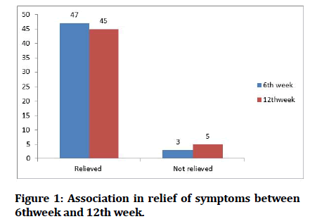
In first week all the 50 had symptoms relieved, in 6th week 47 had symptoms relieved ,in 10th week 45 had symptoms relieved and in 12th week also 45 had symptoms relieved (Tables 1-5) (Figure 1).
| Age in years | Frequency | Percentage |
|---|---|---|
| 21-30 | 4 | 8 |
| 31-40 | 9 | 18 |
| 41-50 | 14 | 28 |
| 51-60 | 23 | 46 |
| Total | 50 | 100 |
| Mean age=47.10 years | ||
| Standard deviation=9.46 | ||
Table 1: Age wise distribution of study participants.
| Sex | Frequency | Percentage |
|---|---|---|
| Male | 12 | 24 |
| Female | 38 | 76 |
| Total | 50 | 100 |
Table 2: Sexwise distribution of study participants.
| Patency | 1st week | 6th week | 10thweek | 12th week |
|---|---|---|---|---|
| Patent | 50 | 47 | 45 | 45 |
| Not patent | 0 | 3 | 5 | 5 |
| Total | 50 | 50 | 50 | 50 |
Table 3: Patency of duct on syringing.
| 1stweek | 6th week | 10th week | 12th week | |
|---|---|---|---|---|
| Relieved | 50 | 47 | 45 | 45 |
| Not relieved | 0 | 3 | 5 | 5 |
| Total | 50 | 50 | 50 | 50 |
Table 4: Relief of symptoms.
| Relieved | Not relieved | Chi square | P.value | |
|---|---|---|---|---|
| 6th week | 47 | 3 | 28.72 | 0.0001 |
| 12thweek | 45 | 5 |
Table 5: Association in relief of symptoms between 6thweekand 12th week.
Figure 1:Association in relief of symptoms between 6thweek and 12th week.
Discussion
Naso-lacrimal duct blockage most commonly presents with epiphora, other symptoms include mucoid or mucopurulent discharge from the eyes and swelling over the medial canthus. Watering is usually unilateral, persisting symptoms may predispose to acute and chronic dacrocystitis if left untreated.
Hence definitive treatment of naso-lacrimal duct blockage is Dacryocystorhinostomy.
Endoscopic Dacryocystorhinostomy is better compared to conventional Dacryocystorhinostomy because of its advantages. There is no scar externally, tissue injury is very minimal and is limited to the fistula area, duration of stay in hospital is less, associated nasal and sinus pathology can be corrected simultaneously. The technique needs expertise in use of endoscopes.
The study was conducted on 50 patients with chronic epiphora with duct obstruction after taking consent from the patient in SBMCH. Patients were examined at outpatient department of Otorhinolaryngology, Sree Balaji Hospital. Examination included ant and post rhinoscopy, diagnostic nasal endoscopy and computed tomography of the Paranasal sinuses to rule out
Any nasal pathology producing nasolacrimal duct obstruction.
To assess any deviation of septum obstructing the view of lacrimal sac area and
Any associated chronic sinusitis / nasal polyps / tumours.
Patients who fulfil the inclusion criteria were selected for the study and all the patients underwent Dacryocystorhinostomy and was evaluated clinically and endoscopically for the subjective and objective relief of symptoms at three months respectively.
The patients were evaluated for systemic conditions like diabetes mellitus and hypertension. The surgical outcomes and the differences of both conventional and endoscopic technique were explained clearly too all patients and a informed written consent was obtained for the surgery
In our study population, majority belong to age group of 51-60 years (46%). About 28% belong to age group of 41-50 years.18% belong to age group of 31-40 years. About 8% belong to age group of 21-30 years. Mean age is 47.10 years. Standard deviations 9.46. Range is from 22-49 years.
In our study 76% were females and 24% were males showing female preponderance to the disease.
Regarding laterality 52% were in the right side,44% in the left side and 4% presented bilaterally. In our study, 52% had right chronic dacryocystitis, 44% had left chronic dacryocystitis and 4%had bilateral chronic dacryocystitis. In our study Bilateral Endoscopic Dacryocystorhinostomy was done for 4%, Right Endoscopic Dacryocystorhinostomy was done for 48% and Left Endoscopic Dacryocystorhinostomy was done for 48%.
For 1stweek, 50 patients had patent duct, in 6th week 47 had patent duct, in 10th week 45 had patent duct an in 12th week also 45 had patent duct. Association in patency between 6th week and 10th week is statistically significant (P value=0.0001) Association in patency between 6th week and 12th week is statistically significant.(P value=0.0001)
In first week all the 50 had symptoms relieved, in 6th week 3 had symptoms, in 10th week 5 had symptoms and in 12th week 5 had symptoms. Association in relief of symptoms between 6thweekand 10th week is statistically significant (P=0.0001). Association in relief of symptoms between 6thweekand 10th week is statistically significant (P=0.0001).
We avoided using silicone tubing but concentrated on exposure of full lacrimal sac, arsuplisation of the sac and the trimming of nasal flap mucosa. With good patient selection, good exposure of lacrimal sac, good surgical technique and good postoperative care we could achieve success rate as high as that of external DCR [2-10].
Conclusion
The conclusion of present study are summarised as follows:
It is common in middle age. Very common in 35-50years age group. Maximum number of cases (46%) was in the fourth decade.
Females (76%) are affected more than males. (4: 1 ratio), it affects both sides commonly.
Treating the concurrent nasal disease appropriately prior to the lacrimal surgery improves the outcome and high septal deviation correction helps to improve results of surgery. Bilateral diseases could be managed simultaneously without much problem.
90% patient had complete relief of symptoms .The success rate achieved by endonasal Dacryocystorhinostomy was as high as that of conventional Dacryocystorhinostomy without the disadvantages of standard conventional approach.
References
- Önerci M. Dacryocystorhinostomy diagnosis and treatment of nasolacrimalcanal obstructions. Rhinology 2002; 40:49–65.
- Gurler B, San I. Long-term follow-up outcomes of non-laser intranasal endoscopic dacryocystorhinostomy: How suitable and useful are conventional surgical instruments? Eur J Ophthalmol 2004; 14:453–460.
- https://journals.lww.com/academicmedicine/citation/1964/09000/system_of_ophthalmology__vol__iii__normal_and.17.aspx
- de la Cuadra-Blanco C, Peces-Pena MD, Merida-Velasco JR. Morphogenesisof the human lacrimal gland. J Anat 2003; 203:531–536.
- https://entokey.com/prenatal-development-of-the-eye-and-its-adnexa/
- Schaeffer JP. The genesis and development of the nasolacrimal passages inman. Am J Anat 1912; 13:1–23.
- Jones LT, Wobig JL. Congenital anomalies of the lacrimal system. In: Surgery of the eyelids and lacrimal system. Birmingham, AL: Aesculapius 1976; 157–173.
- Hurwitz JJ. Embryology of the lacrimal drainage system. In: Hurwitz JJ,ed. The lacrimal system. Philadelphia: Lippincott-Raven 1996; 9–13.
- Petersen RA, Robb RM. The natural course of congenital obstruction of the nasolacrimal duct. J Pediatr Ophthalmol Strabismus 1978; 15:246–250.
- Fooks OO. Dacryocystitis in infancy. Br J Ophthalmol 1962; 46:422.
Author Info
Gopi Ayyasami, Mohan Kumar J* and Jaya Preetha
Department of ENT, Sree Balaji Medical College and Hospital, IndiaCitation: Gopi Ayyasami , Mohan Kumar J, Jaya Preetha, Role of Endoscopic Dacryocystorhinostomy in Patients with Chronic Epiphora, J Res Med Dent Sci, 2021, 9(11): 253-256
Received: 06-Oct-2021 Accepted: 15-Nov-2021

