Research - (2019) Volume 7, Issue 4
The Impact of Selected Fluoride Materials and Nd: YAG LASER on Dentine (In Vitro Study)
Khamaal IM Al-Hasnawi* and Nada Jafer MH Radhi
*Correspondence: Khamaal IM Al-Hasnawi, Pedodontic and Preventive Dentistry Department, College of Dentistry, University of Baghdad, Baghdad, Iraq, Email:
Abstract
Background: Topical fluoride use result in re harden or arrest lesions of dental caries in dentine. Silver diamine fluoride (SDF) can be employed to inhibit and arrest dental caries. The use of laser in caries prevention was suggested by used Neodymium doped yttrium-aluminium-garnet (Nd: YAG) laser as an adjunct to conventional caries prevention therapies.
Aim: The aim of this research was to investigate the effect of selected topical fluoride materials, acidulated phosphate fluoride (APF), Silver diamine fluoride (SDF), Stannous fluoride (SnF2) with or without Nd-YAG laser application on Fluoride uptake of dentine, in vitro.
Materials and methods: The samples in this study include 55 maxillary first premolar teeth, distributed into 11 groups, and each group involved of five specimens. Before the surface treatments, all the specimens in the 11 groups underwent a pH cycling procedure to induced caries like lesion in dentine specimen for five days, and then 2 days in remineralization solutions. Fluoride measurements were accomplished by an ion-selective electrode.
Results: Results presented after treatment with different agents, a marked increase in fluoride uptake for the groups treated with SDF+Laser, SnF2, Laser+SnF2, Laser+APF. While a severe increase in fluoride uptake for the group treated with silver diamine fluoride (SDF) alone. Conclusion: Treatment of dentine surface with Silver diamine fluoride alone gave the best result regarding as topical fluoride application.
Keywords
Nd: YAG laser, Silver diamine fluoride, Acidulated phosphate fluoride, Stannous fluoride
Introduction
Dental caries refers to the localized chemical termination of dental hard tissue that result from acidic by products of dental plaque’s (biofilm) metabolic processes that cover an affected tooth surface [1]. Changes occur in both enamel and dentin but the progression of caries is not the same. In enamel, dissolution occur in highly mineralised tissue as a result of bacterial acid [2], whereas that in dentine includes both demineralization of mineral and organic matrix dissolution of the type I collagen fiber system [3]. Earlier, the damage of the dentine organic matrix was measured to be mostly occurring because of the bacterial collagenases action, whereas some researches have shown that collagen is likely to be ruined by matrix metalloproteinases [4], they are triggered by low pH within the carious dentine [5].
Fluoride are chief assistant in the prevention of dental caries, however it takings time for depositioning as it requires about 3ppm change the balance from net demineralization into net re-mineralization [6]. The higher intake of fluoride in the period of tooth development increases the amount of fluoride in enamel, thus, raising the amount of acid resistance and this formed the hypothesis. Due to this, synthetic fluoridation of water for drinking turned to be the best solution and there was need to take fluoride systemically [7]. Majority of the fluoridation systems offer less concentrations of fluoride ions in saliva, therefore, the plaque fluid experiences a topical impact. Fluoride stops the caries mostly through enamel remineralization resulting in fluorapatite formation. The ions of fluoride minimize demineralization of tooth enamel, and raise the remineralization rate of cavities in the initial stages [8].
Topical fluorides are delivered to teeth at exposed surfaces, at high amounts for a local defensive impact to make the teeth extra resistant to caries. Topical fluorides are applied to the teeth directly and are most effective when delivered at very low doses several times a day [9]. The fluoride compound formed for topical application in the dental department in 1950s included SnF2 (stannous fluoride), the reaction between enamel and stannous fluoride is exceptional in the manner that all the reactants anion (fluoride) and cation (stannous) chemically act in response with enamel constituents [10]. Acidulated phosphate fluoride (APF) was brought in 1960s inform of a gel and solution. Skilfully used acidulated phosphate fluoride comprises of 12,300 ppm fluoride (1.23% fluoride) as sodium fluoride at pH 3. The solution was created according to the researches premeditated to hasten formation of enamel fluorapatite, reduce the formation of calcium fluoride, raise fluoride surface absorption, and demineralization prevention [11]. Silver diamine fluoride is not just a meek salt of ammonium, fluoride, and silver ions, but it a varied thick-metal halide multifaceted combination. Ammonia has the ability to maintain the solution at a frequent concentration in a long period [12]. Therefore, the joint impact of fluorides and silver has been theorized to possess the potential to combat caries development and avert emergent of fresh caries instantaneously [13].
Laser irradiation can be spread, absorbed, diffused or reflected into dental tissues Depending on tissue features and laser wavelength [14]. The neodymium yttriumaluminum- garnet (Nd:YAG) laser is a solid-state apparatus that produce light in the near infrared region of the spectrum at 1064 nm. Lasing has an antibacterial action on enamel and dentine through the vaporization of cariogenic bacteria both on the surface and in the subsurface configuration [15]. Studies in-vitro shown that Nd:YAG radiation of a dentine or enamel surface decreases its permeability [16]. As far as there was no Iraqi study directed to explore the impact of silver diamine fluoride with or without Nd-YAG laser, this study was conducted.
Materials and Methods
The samples in this study comprised of 55 maxillary first premolar teeth. Samples were erratically distributed into 11 groups, and each group involved of five specimens for fluoride uptake test. Teeth had been distributed into groups consistent with the type of treatment:
Group 1: Control teeth with neither laser nor silver diamine fluoride, acidulated phosphate fluoride and Stannous fluoride treatment.
Group 2: Silver diamine fluoride-treated teeth.
Group 3: Acidulated phosphate fluoride-tread teeth.
Group 4: Teeth treated with Stannous fluoride.
Group 5: Nd-YAG laser-treated teeth (Figure 1).
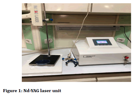
Figure 1. Nd-YAG laser unit
Group 6: Teeth treated with laser then silver diamine fluoride.
Group 7: Teeth treated with silver diamine fluoride solution then laser.
Group 8: Teeth treated with laser then Stannous fluoride.
Group 9: Teeth treated with Stannous fluoride solution then laser.
Group 10: Teeth treated with acidulated phosphate fluoride then laser.
Group 11: Teeth treated with laser then acidulated phosphate fluoride.
The buccal surface of the crowns was selected for this experiment. The tooth was sliced by a high speed turbine hand piece with flow of de-ionized water to form square slabs (the enamel window technique) which described by Grenby et al. [17], at the center of the image line extending to the cement-enamel-junction (CEJ) from the uppermost cusp, the primary cut was completed mesiodistally via the tip of the buccal cusp in order to access the buccal surface dissection whereas holding the root and there was removal of coronal dentine alongside carbide round burs slowly using a light with the used of magnifying eyeglass. Serving of the sample was at about a mid-third of the buccal surface as seen in (Figure 2). Then on buccal side was cautiously cut off to expose a flat dentine surface. 2.5 by 2.5 mm window in surface in the tooth block (Figure 3). Tooth block surface were coated by acid resilient nail paint, except the dentine window. The tooth blocks were labeled individually on one of the painted flat surface.
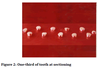
Figure 2. One-third of tooth at sectioning
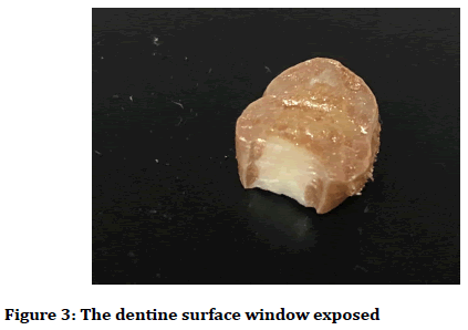
Figure 3. The dentine surface window exposed
Before the surface treatments, all the specimens in the 11 groups underwent a pH cycling procedure to induced caries like lesion in dentine specimen for five days [18,19], and then 2 day in remineralizing solutions. The demineralizing and remineralizing solutions were prepared similarly to that in a study by Jeon et al. [20].
After each dentine specimen treatment, it was etched with 10 μl of 2M HCl for 10 second. The liquid was then collected using of paper points, subsequently transferred to 5 ml water solution and TISAB III 0.5 ml (0.4% EDTA and 1.0 M acetate buffer 1 M NaCL, pH 5.0) that necessary to immerging fluoride electrode adequately [21].
Fluoride quantities were carried out through the application of an ion-selective electrode (WTW) GmbH (Germany) combination of fluoride electrode F 800 and F 500 (Figure 4), to establish the concentration of fluoride ions within dentine sections by the potential in standard solutions’ millivolt (mV) were openly estimated using the electrode then calibrated with the application of a concentration scale or through plotting calibration graph and created on a semi-logarithmic standard paper through drawing long axis (concentration) versus linear axis (the millivolt interpretations).
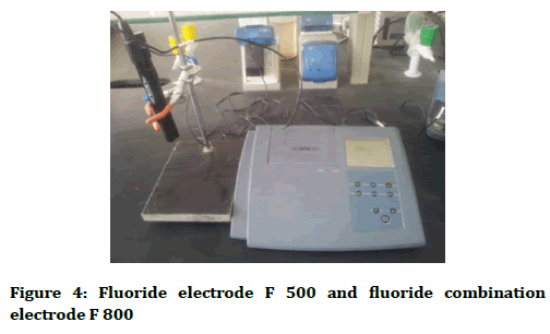
Figure 4. Fluoride electrode F 500 and fluoride combination electrode F 800
Statistical analysis
Description, analysis and presentation of data were carried out with the application of SPSS (Statistical Package for Social Science) version 21. The statistical analyses may be grouped into 2 classes:
Descriptive analysis
• Mean and standard deviation for quantitative variable.
• Graphs: 1-Simple and means plot with error bars with 95% Confidence Intervals (CI); 2-Box-plot bars.
Inferential statistics
• Shapiro-Wilk test: It measures normality distribution of variable.
• Levene test: It analyses the homogeneity of variance among groups.
• One-Way-ANOVA (Analysis of Variance): Parametric test determines and find difference between K independent samples with Tukey HSD (Tukey Honestly Significant Difference), Dunnett's T3 and Dunnett t (2- sided) post hoc tests. Significance levels as: Significant p<0.05, Non-significant p>0.05, and highly significant p<0.01.
Results
The mean values of fluoride uptake of dentine surfaces afterward synthetic caries-like lesion creation control (Con) and following treatment with silver diamine fluoride (SDF), Acidulated phosphate fluoride (APF), Stannous fluoride (SnF2), Nd-YAG Laser, Nd-YAG Laser +SDF, SDF+Nd-YAG Laser, Nd-YAG Laser+SnF2, SnF2+ Nd- YAG Laser, APF+Nd-YAG Laser, Nd-YAG Laser+APF are seen in Table 1 and Figure 5.
| Groups | Mean | SD | Levene | p-value | F | p-value |
|---|---|---|---|---|---|---|
| Con | 0.002 | 0.002 | 6.009 | 0 | 40.58 | 0.000** |
| SDF | 0.08 | 0.004 | ||||
| APF | 0.028 | 0.005 | ||||
| SnF2 | 0.042 | 0.005 | ||||
| Laser | 0.013 | 0.006 | ||||
| L+SDF | 0.032 | 0.006 | ||||
| SDF+L | 0.045 | 0.002 | ||||
| L+SnF2 | 0.037 | 0.008 | ||||
| SnF2+L | 0.026 | 0.013 | ||||
| APF+L | 0.026 | 0.013 | ||||
| L+APF | 0.037 | 0.003 |
Table 1: Fluoride uptake (men and standard deviation) of dentine surfaces for selected materials.
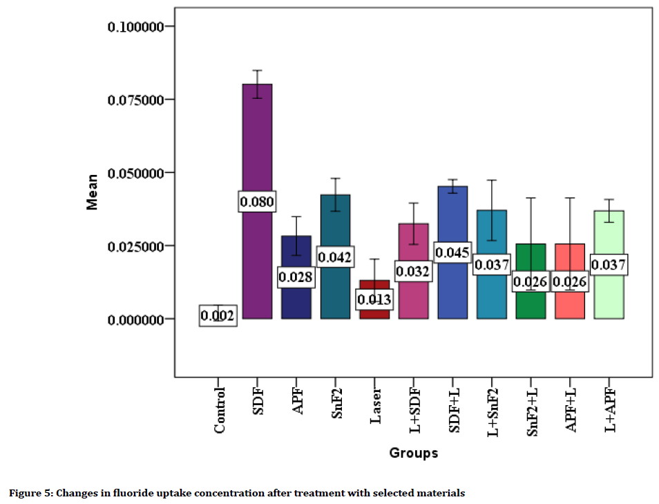
Figure 5. Changes in fluoride uptake concentration after treatment with selected materials
Multiple comparisons of fluoride uptake for all groups could be seen in Table 2, it was found a statistically highly significant difference in fluoride uptake for the control group when compared with group treated with SDF, APF, SnF2, Laser+SDF, SDF+Laser, Laser+SnF2, Laser+APF (p<0.01). Non-significant differences were shown among control group and (Laser, SnF2+Laser, APF+Laser) groups (p>0.05).
| (I) Group | (J) Group | ||||||||||
|---|---|---|---|---|---|---|---|---|---|---|---|
| SDF | APF | SnF2 | Laser | L+SDF | SDF+L | L+SnF2 | SnF2+L | APF+L | L+APF | ||
| Con | Mean Difference (I-J) | -0.078 | -0.026 | -0.04 | -0.011 | -0.03 | -0.043 | -0.035 | -0.024 | -0.024 | -0.035 |
| p value | 0.000** | 0.003** | 0.000** | 0.166 | 0.002** | 0.000** | 0.008** | 0.181 | 0.181 | 0.000** | |
| SDF | Mean Difference (I-J) | 0.052 | 0.038 | 0.067 | 0.048 | 0.035 | 0.043 | 0.055 | 0.055 | 0.043 | |
| p value | 0.000** | 0.000** | 0.000** | 0.000** | 0.000** | 0.002** | 0.007** | 0.007** | 0.000** | ||
| APF | Mean Difference (I-J) | -0.014 | 0.015 | -0.004 | -0.017 | -0.009 | 0.003 | 0.003 | -0.009 | ||
| p value | 0.059 | 0.075 | 0.998 | 0.022* | 0.825 | 1 | 1 | 0.326 | |||
| SnF2 | Mean Difference (I-J) | 0.029 | 0.01 | -0.003 | 0.005 | 0.017 | 0.017 | 0.005 | |||
| p value | 0.001** | 0.331 | 0.988 | 0.995 | 0.469 | 0.469 | 0.72 | ||||
| Laser | Mean Difference (I-J) | -0.019- | -0.032 | -0.024 | -0.012 | -0.012 | -0.024 | ||||
| p value | 0.022* | 0.002** | 0.030* | 0.815 | 0.815 | 0.005** | |||||
| L+SDF | Mean Difference (I-J) | -0.013 | -0.005 | 0.007 | 0.007 | -0.004 | |||||
| p value | 0.092 | 1 | 0.999 | 0.999 | 0.968 | ||||||
| SDF +L | Mean Difference (I-J) | 0.008 | 0.02 | 0.02 | 0.008 | ||||||
| p value | 0.739 | 0.3 | 0.3 | 0.042* | |||||||
| L+SnF2 | Mean Difference (I-J) | 0.011 | 0.011 | 0 | |||||||
| p value | 0.933 | 0.933 | 1 | ||||||||
| SnF2+L | Mean Difference (I-J) | 0 | -0.011 | ||||||||
| p value | 1 | 0.827 | |||||||||
| APF+L | Mean Difference (I-J) | -0.011 | |||||||||
| p value | 0.827 | ||||||||||
Table 2: Multiple comparisons of Fluoride uptake of dentine surfaces for selected materials for all groups by using Dunnett T3 test
A statistically highly significant differences were found when comparing the group treated with SDF with all groups (p<0.01). The group treated with APF showed a statistically significant difference in comparison with the group treated with SDF+L (p<0.05) while, non-significant differences were shown with the others. The same for the group treated with SnF2 showed a statistically highly significant difference when compared with the group treated with Laser (p<0.01) while, non-significant differences were shown with the other groups.
The group treated with Laser showed a statistically significant difference when compared with the groups treated with (Laser+ SDF, Laser+SnF2) (p<0.05) whereas, a highly significant difference when compared with the group treated with (SDF+Laser, Laser+APF) (p<0.01) and non-significant differences were shown among other groups (p>0.05). Whereas, a statistically significant difference was shown between the SDF+Laser group when compared with Laser+APF group (p<0.05), while non-significant differences were shown among other groups (p>0.05). The groups treated with (Laser+SDF, SnF2+Laser, Laser+SnF2, APF+Laser) were shown nonsignificant differences among other groups (p>0.05).
Discussion
Fluoride uptake measurement was applied in this study to reflect exactly the degree of remineralization of the lesion. This was accomplished by Ion-selective electrode method that gave successful results by a previous study [22]. The measurement of fluoride uptake was doing for sound dentine, after subjected to demineralization and remineralizing sequence (pH cycling) for five days [23] and subsequent treatment with the selected materials, then remineralization for 2 days.
Result showed that there was a marked rise in uptake of fluoride by dentine after treatment stage with selected topical fluoride material alone or with laser as compared to fluoride concentration of control teeth for all groups. After treatment with selected material, a marked increase in fluoride uptake for the groups treated with APF, SnF2, Laser+SDF, SDF+Laser, Laser+SnF2, Laser+APF. While a sharp increase in fluoride uptake for the group treated with silver diamine fluoride (SDF) alone.
After applying silver diamine fluoride on a caries surface, the squamous layer of silver-protein combines emerges, raising the enzymatic digestion and acid dissolution resistance [24]. The consequences of indicated laboratory researches usually approve that silver diamine fluoride concentration of 38 percent is very operative when it comes to preventing collagenase action and deterrence collagen destruction as compared to less concentration [5,25]. Fluoride and Silver ions infiltrate ~25 microns to enamel, and 50-200 microns to dentin. Silver diamine fluoride arrested lesions are 150 microns in thickness [26].
The great level of fluoride taken up by enamel and dentine treated with APF gel may describe its higher ability in decreasing equally caries and erosion when associated to NaF gel [27].
The process for the defensive influence of SnF2 alongside erosion can be described by the obstructive diffusion movement [28]. The dentinal superficial is protected by a film having the reactive produces of SnF2 and hydroxyapatite, as salts of Sn2OHPO4, Ca (SnF3)2, Sn3F3PO4, or CaF2, that can eliminate obvious dentinal tubules. On the other hand, in compare to previous studies, [29,30] SnF2 unaccompanied was incapable to decrease the demineralization and loss of mineral from dentine.
The Nd:YAG laser radiation reduced the quantity of carbonate of dentin when related to the phosphate quantity, representing the happening of carbonate damage because of laser radiation. The carbonate radical can standby the hydroxyl or phosphate radicals in the natural apatite crystal, creating a fewer constant and less solvable stage [31]. On the other hand, opposing consequences are also detected, since it was informed that this wavelength of laser cannot suggestion any extra assistance when related to fluoride inaction alone [32] that is in agreement with present study of laser group statistically non-significant difference in fluoride uptake values with control group.
A study showed that low-energy sub-ablative Nd:YAG laser radiation subsequent SDF usage, enlarged fluoride installation and altered hydroxyapatite to fluoridated hydroxyapatite [33], the description is that once SDF can source the stain of demineralized dentine (the result is not important on intact dentine), improving the engagement the energy of Nd:YAG laser as the Nd:YAG laser are called pigment dependence laser that mostly cooperate with stained tissues as melanin, and are merely a little absorbed by water [34].
For the group treated with SnF2 or APF followed by laser, the mean values of fluoride uptake have revealed non-significant difference as compared with control group, approved with the authors described that this would show laser action has no result on the hypersensitivity of the tooth and demineralization of the dentine as SnF2 or APF disguised the laser effect such as a topical fluoride use before it [35,36].
For the groups treated with Nd:YAG laser then one of these materials (APF, SDF, SnF2) result shown that the mean values of fluoride uptake increased, this may be recognized to the fact that laser parameters in the present study were used, they were enough to encourage the melting and recrystallized to greater apatite crystals comprising fewer carbonate during laser radiation of the dentin surface, moreover, they were revealed to be harmless for pulp liveliness [32,37,38].
Conclusion
A highly significant difference in fluoride uptake was recorded among groups as comparing with control group. The distinct increase in fluoride uptake for the groups treated with SDF+ Laser, SnF2, Laser+ SnF2, Laser +APF. While a severe increase in fluoride uptake for the group treated with silver diamine fluoride (SDF) alone. Treatment of dentine surface with Silver diamine fluoride alone gave the best result regarding as topical fluoride application, so such treatment could be considered as a method of preventing demineralization and encouraging remineralization of dentine.
Conflict of Interest
The authors declare that there is no conflict of interest regarding the publication of this manuscript.
References
- Selwitz RH, Ismail AI, Pitts NB. Dental caries. Lancet 2007; 369:51-9.
- Mei ML, Ito L, Cao Y, et al. Inhibitory effect of silver diamine fluoride on dentine demineralisation and collagen degradation. J Dent 2013; 41:809-17.
- Horst JA, Ellenikiotis H, Milgrom PL. UCSF protocol for caries arrest using silver diamine fluoride: Rationale, indications and consent. J Calif Dent Assoc 2016; 44:16-28.
- Chaussain-Miller C, Fioretti F, Goldberg M, et al. The role of matrix metalloproteinases (MMPs) in human caries. J Dent Res 2006; 85:22-32.
- Mei ML, Li QL, Chu CH, et al. The inhibitory effects of silver diamine fluoride at different concentrations on matrix metalloproteinases. Dental Mater 2012; 28:903-08.
- Summit JB, Robbins WJ, Schwartz RS. Fundamentals of operative dentistry. A contemporary approach, 2nd ed. Chicago, IL: Quintessence Publishing 2001; 377-85.
- Fejerskov O. Changing paradigms in concepts on dental caries: Consequences for oral health care. Caries Res 2004; 38:182-91.
- Pizzo G, Piscopo MR, Pizzo I, et al. Community water fluoridation and caries prevention: A critical review. Clin Oral Investig 2007; 11:189-93.
- Ijaz S, Marinho VCC, Croucher R, et al. Professionally applied fluoride paint-on solutions for the control of dental caries in children and adolescents. Cochrane Database Syst Rev 2010; 2.
- Muhler JC, Boyd TM, Van Huysen G. Effects of fluorides and other compounds on the solubility of enamel, dentin, and tricalcium phosphate in dilute acids. J Dent Res 1950; 29:182-93.
- Chambers MS, Mellberg JR, Keene HJ, et al. Clinical evaluation of the intraoral fluoride releasing system in radiation-induced xerostomic subjects. Part 1: Fluorides. Oral Oncol 2006; 42:934-45.
- Mei ML, Lo EC, Chu CH. Clinical use of silver diamine fluoride in dental treatment. Compend Contin Educ Dent 2006; 37:93-8.
- Rosenblatt A, Stamford T, Niederman R. Silver diamine fluoride: A caries “silver-fluoride bullet”. J Dent Res 2009; 88:116-25.
- Ana PA, Bachmann L, Zezell DM. Lasers effects on enamel for caries prevention. Laser Physics 2006; 6:865-75.
- Fuller TA. Physical considerations of surgical lasers. In: Clayman L, editor: Oral Maxillofac Surg Clin North Am 1997; 9:1-9.
- Tagomori S, Iwase T. Ultrastructural change of enamel exposed to a normal pulsed Nd-YAG laser. Caries Res 1995; 29:513-20.
- Grenby TH, Bull JM. Chemical studies of the protective action of phosphate compounds against the demineralization of human dental enamel in vitro. Caries Res 1980; 14:210-20.
- Argenta RM, Tabchoury CP, Cury JA. A modified pH-cycling model to evaluate fluoride effect on enamel demineralization. Brazilian Oral Research 2003; 17:241-6.
- Fahad AH, Radhi NJ. Effect of casein phosphopeptide-amorphous calcium phosphate on the microhardness and microscopic features of the sound enamel and initial caries-like lesion of permanent teeth, compared to fluoridated agents. J Bagh College Dentistry 2012; 24:114-120.
- Jeon RJ, Hellen A, Matvienko A, et al. Experimental investigation of demineralization and remineralization of human teeth using infrared photothermal radiometry and modulated luminescence. In Photons Plus Ultrasound: Imaging and Sensing 2008: The Ninth Conference on Biomedical Thermoacoustics, Optoacoustics, and Acousto-optics. International Society for Optics and Photonics 2008; 6856:68560B.
- Orion instruction manual. Thermo Fisher Scientific protected by US Patent 6, 350, 376. Orion 2007.
- AL-Hasnawi KI, Al-Obaidi WA. Effect of Nd-YAG laser irradiation on fluoride uptake by tooth enamel surface (in vitro). J Bagh College Dentistry 2014; 26:154-58.
- Featherstone J, Tencate M, Shariati J, et al. Comparison of artificial caries like lesion by quantitative micro radiography and microhardness profiles. Caries Res 1986; 17:385-91.
- Mei ML, Chu CH, Lo EC, et al. Fluoride and silver concentrations of silver diamine fluoride solutions for dental use. Int J Paediatr Dent 2012; 23:279-285.
- Mei ML, Ito L, Cao Y, et al. The inhibitory effects of silver diamine fluorides on cysteine cathepsins. J Dent 2014; 42:329-335.
- Chu CH, Lo EC. Promoting caries arrest in children with silver diamine fluoride: A review. Oral Health Prev Dent 2008; 6:315-321.
- Jones L, Lekkas D, Hunt D, et al. Studies on dental erosion: An in vivo-in vitro model of endogenous erosion – its application to testing protection by fluoride gel application. Aust Dent J 2002; 47:304-308.
- Tepper SA, Zehnder M, Pajarola GF, et al. Increased fluoride uptake and acid resistance by CO2 laser irradiation through topically applied fluoride on human enamel in vitro. J Dent 2004; 32:635-41.
- Willumsen T, Ogaard B, Hansen BF, et al. Effects from pretreatment of stannous fluoride versus sodium fluoride on enamel exposed to 0.1 M or 0.01 M hydrochloric acid. Acta Odontol Scand 2004; 62:278-81.
- Young A, Thrane PS, Saxegaard E, et al. Effect of stannous fluoride toothpaste on erosion-like lesions: An in vivo study. Eur J Oral Sci 2006; 114:180-83.
- Robinson C, Shore RC, Brookes SJ, et al. The chemistry of enamel caries. Crit Rev Oral Biol Med 2000; 11:481-95.
- Zezell DM, Boari HG, Ana PA, et al. Nd:YAG laser in caries prevention: A clinical trial. Lasers Surg Med 2009; 41:31-5.
- Liu Y, Hsu CY, Teo CM, et al. Subablative Er: YAG laser effect on enamel demineralization. Caries Res 2013; 47:63-8.
- Zhang C, Kimura Y, Matsumoto K, et al. Effects of pulsed Nd: YAG laser irradiation on root canal wall dentin with different laser initiators. J Endod 1998; 24:352-55.
- Sicilia A, Cuesta – Frechoso S, Sua´rez A, et al. Immediateefficacy of diode laser application in the treatment of dentine hypersensitivity in periodontal maintenance pa-tients: A randomized clinical trial. J Clin Periodontol 2009; 36:650-60.
- Lee R, Chan KH, Jew J, et al. Synergistic effect of fluoride and laser irradiation for the inhibition of the demineralization of dental enamel. Proc SPIE 2017; 10044.
- Celik EU, Ergucu Z, Turkun LS, et al. Effect of different laser devices on the composition and micro-hardness of dentin. Oper Dent 2008; 33:496-501.
- Passos VF, Melo MS, Silva FC, et al. Effects of diode laser therapy and stannous fluoride on dentin resistance under different erosive acid attacks. Photomed Laser Surg 2014; 32:146-15.
Author Info
Khamaal IM Al-Hasnawi* and Nada Jafer MH Radhi
Pedodontic and Preventive Dentistry Department, College of Dentistry, University of Baghdad, Baghdad, IraqCitation: Khamaal IM Al-Hasnawi, Nada Jafer MH Radhi, The Impact of Selected Fluoride Materials and Nd: YAG LASER on Dentine (In Vitro Study), J Res Med Dent Sci, 2019, 7(4): 1-7.
Received: 30-May-2019 Accepted: 19-Jul-2019
