Mini Review - (2021) Volume 9, Issue 12
To Evaluate the Total Tooth Length of the MandibularAnteriors Treated by Extraction of all 4first Premolar in Class I Bimaxillary Protrusion Cases Using CBCT - A Comparative
Hima Shwetha1*, Dharmesh H2 and Bharathi3
*Correspondence: Hima Shwetha, Dept. Of Orthodontics and Dentofacial Orthopaedics, Rajarajeswari Dental College and hospital, Bangalore, India, Email:
Abstract
Aim: To evaluate the changes in the total tooth length during lower incisor retraction in Bimaxillary protrusion cases (Class I)using Cone Beam Computed tomographic images. Methodology: Ten patients undergoing orthodontic treatment with MBT 3M (Abzil) - 0.022 slot appliance were selected. Change in periapical region was measured by comparing the pre-treatment and post-retraction CBCT of the Mandibular anterior region. ONDEMAND Software was used for image processing and analysis to evaluate the changes in the Total Tooth Length [TTL] during lower incisor retraction in class I Bimaxillary protrusion cases. Result: Total tooth length was reduced compared to the pretreatment CBCT images. The mean Total tooth length [TTL] Pretreatment [T0] was 18.38 mm and Post-treatment [T1] was 17.38 mm; the difference value of mean Total Tooth Length [TTL] was 1.0 mm (p value = 0.02). Conclusion: The study showed that there was a decrease in the total tooth length [TTL].
Keywords
Cone Beam Computed Tomography, Total tooth length, SS-Stainless steel wiresIntroduction
Asian orthodontic patients are commonly seen with Bialveolar protrusion. The conventional treatment is extraction of the first permanent premolars and retraction of the anterior teeth with Maximum Anchorage (Critical Anchorage). Iatrogenic sequelae such as Root resorption, Alveolar bone loss, Dehiscence, Fenestration and Gingival recession will be noticed upon excessive retraction of the anterior teeth.
Several limitations have been found in Conventional two-dimensional (2D) lateral cephalograms in investigating the changes in the alveolar bone and roots, especially in the anterior region, in midsagittal projection. Qualitative and quantitative evaluation can be made with the advent of cone-beam computed tomography (CBCT) to evaluate the length, height, alveolar bone thickness and the root thickness. The Patient’s hard and soft tissue can be evaluated in three dimensions with the advent of CBCT scan. The three-dimensional images have been found to be effective for orthodontic purposes in both accuracy and reliability.
Objectives
To compare the Pre-treatment [T0] and Post-retraction [T1] Total Tooth Length of the mandibular anteriors using CBCT. Treated by extraction of all 4 first premolar in Bimaxillary protrusion cases.
Material and method
Ten patients with Bimaxillary protrusion between the age 15-30 yrs were selected who desired to undergo orthodontic treatment with Preadjusted edgewise appliance [PEA], MBT 3M Initial leveling and aligning was achieved using 0.016NiTi archwire. Later, the wire sequencing followed was Niti Niti, Niti and finally retraction was carried out using (Stainless Steel) archwire along with soldered hooks incorporated in between the mandibular laterals and mandibular canines as the Centre of resistance [CR] of lower anteriors lie between the mandibular lateral incisors and the canines.
Assessment was done in the retracted area using Pretreatment (T0) and Post retraction (T1) CBCT images for evaluation of the changes in the Total Tooth Length [TTL] during lower incisor retraction in class I Bimaxillary protrusion cases. Field of view was 75mm×145mm with a voxel size of 0.25mm, 90 kvp and 12mA and exposure time of 15 seconds. Ondemand software was used for image processing and analysis with screen resolution of 1920×1200 pixels and 64-bit color. Measurements on scan was made using Ondemand software from incisal edge to root apex 1. Single investigator determined all measurements.
Results
Evaluation of Total Tooth Length [TTL] treated by extraction of all 4 first premolar in Bimaxillary protrusion cases during pre-retraction [T0] and post retraction [T1] using computed tomography images.
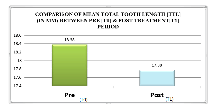
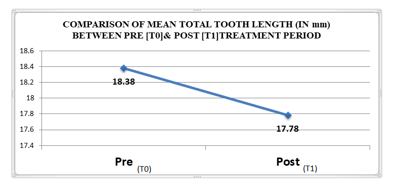
The test results demonstrate that the mean total tooth length [TTL] Pretreatment [T0] was 18.38 mm and Post-treatment [T1] was 17.38 mm; the difference value of mean Total Tooth Length [TTL] with statistically significant results From the above results it can be concluded that the Total Tooth Length [TTL] is decreased in Post treatment [T1] compared to Pretreatment [T0] by 1mm (Graph 1&2).
Graph 1: Bar graph representing the comparison of total tooth length [ttl] cases between pre [t0] & post treatment [t1]
Graph 1: comparison of total tooth length[ttl] cases between pre [t0] & post treatment [t1]
Discussion
This study revealed the changes in the total tooth length in lower incisor region and to know its relation with displacement of root apex and inclination changes in mandibular incisors during retraction of anteriors.
Hyo Won Ahn, Sung Chung Moon, Seug Hak Baek conducted a study on Morphometric evaluation of changes in the tooth length and root length of maxillary anterior teeth before and after en mass retraction. The aim of the study was to evaluate dentoalveolar protrusion (CI-DAP) and were treated by extraction of the first premolars and EMRMA. Using three-dimensional CBCT taken before treatment and after space closure. Tooth length (TL), Root length (RL), Root area (RA), and prevalence of dehiscence (PD) were measured at the cervical, middle, and apical levels were measured. The palatal side showed more PD in the cervical area than did the labial side. Significant reduction in the tooth length was seen especially in maxillary lateral incisors and root resorption occurred in MXAT (RL and RA, all P<0.01).
Simplicio H, da Silva JS, Caldas SG, dos Santos-Pinto A conducted a study on External apical root resorption and changes in the tooth length and its inclination in retracted incisors5.The objective of the study was to evaluate the occurrence of external apical root resorption (EARR) in the incisors after anterior retraction in corrective orthodontic treatment with extraction of first premolar and whether it was related with the type of root apex movement and change in tooth length and its inclination. Pre- and post-incisal retraction of CBCT images and lateral cephalometric radiographs established the relationship between EARR, along with the vertical and total length of the tooth from cervical area to root apex was measured. It was observed that after the retraction stage, EARR occurred in all evaluated incisors, but it was more significant (P < .05) in the mandibular right lateral incisor. It was concluded that the EARR occurred upon retraction and there was a significant reduction in the total tooth length measurement in the mandibular anteriors.
Yang Han, Xiao Guang Li conducted a study on Cone-beam computed tomography digital for measuring the inclination and total tooth length of maxillary anterior teeth upon retraction in bimaxillary protrusion cases6. The aim was to determine the total tooth length, the distance between the cement-enamel junction (CEJ) and root apex, the inclination angle of the long axis of maxillary anterior teeth by using cone- beam computed tomography (CBCT). The digital measurements taken from the widest point on the labiolingual root at level 4 mm below the CEJ, the radiological tooth apex, and the angle between long axis of the teeth, mid-sagittal planes of maxillary incisors. There was significant decrease in total tooth length in maxillary central incisor (left) and maxillary lateral incisor.
Conclusion
It was concluded the Total Tooth Length [TTL] was decreased when compared with the Pretreatment [T0] and Postreatment [T1] CBCT images. The mean Total tooth length [TTL] Pretreatment [T0] was 18.38 mm and Post-treatment [T1] was 17.38 mm; the difference value of mean Total Tooth Length [TTL] was 1.0 (p value=0.02)* with statistically significant results. This study reveals that upon mandibular anteriors retraction in Bimaxillary protrusion cases there was a decrease in the Total Tooth Length [TTL].
References
- Yodthong N, Charoemratrote C, Leethanakul C, et al. Factors related to alveolar bone thickness during upper incisor retraction. Ang Ortho 2012; 83:394-401.
- Yu Lan Wan, Houi Xi Kou. Changes in root and alveolar bone before and after treatment by retracting the upper incisors. West china J stomat 2018; 36:638-645.
- Hyo Won Ahn, Sung Chul Moon. Morphometric evaluation of changes in the tooth length and roots of the maxillary anterior teeth before and after en masse retraction using cone-beam computed tomography. Angle orthod 2013; 83:212-21.
- Kyoung Won Kim, Sung Jin Kim. Apical root displacement is a critical risk factor for apical root resorption after orthodontic tooth movement. Angle orthod 2018; 88:740-747.
- Richard J Smith, Charles J Burstone. Mechanics of tooth movement. Am J Orthod 1980; 77:396-409
- Yaqi Deng, Yannan Sun, Tianmin, et al. Evaluation of root resorption after comprehensive orthodontic treatment using cone beam computed tomography.Oral Health 2018; 18:116
Author Info
Hima Shwetha1*, Dharmesh H2 and Bharathi3
1Dept. Of Orthodontics and Dentofacial Orthopaedics, Rajarajeswari Dental College and hospital, Bangalore, India2Dept. of Orthodontics and Dentofacial Orthopaedics, Rajarajeswari Dental College and hospital, Bangalore, India
3Dept. of Orthodontics and Dentofacial Orthopaedics, Rajarajeswari Dental College and hospital, Bangalore, India
Citation: Dr.Hima Shwetha, Dharmesh H, Dr.Bharathi.To Evaluate the Total Tooth Length of the Mandibular Anteriors Treated by Extraction of all 4first Premolar in Class I Bimaxillary Protrusion Cases Using CBCT - A Comparative , J Res Med Dent Sci, 2021, 10(9): 1-3
Received: 01-Dec-2021 Accepted: 15-Dec-2021 Published: 22-Dec-2021


