Research - (2021) Volume 9, Issue 2
Visibility of Mandibular Canal on CBCT Cross-Sectional Images in Comparison with Panoramic Radiograph (Retrospective Study)
Resha Jameel1*, Areej Ahmed Najm1 and Firas Abdulameer Farhan2
*Correspondence: Resha Jameel, Department of Oral and Maxillofacial Radiology, Collage of Dentistry, University of Baghdad, Iraq, Email:
Abstract
Detection of mandibular canal (MC) is an important requirement for different surgical procedures such as implant placement, surgical removal of pathological lesion and even to tooth extraction. Cone beam computed tomography (CBCT) represents an advance imaging technology, but distinguishing the MC from surrounding structures may persists a difficult matter. Purpose: The aim of this study was to assess the visibility of MC in different regions on CBCT cross-sectional images and compare with OPG images. Materials and methods: in this retrospective study 400 hemi mandible (100 males and 100 females) were examined by CBCT cross section and compared to digital panoramic images. the images were evaluated and the mandibular canal visibility was assessed in four region (second premolar, first molar, second molar and distal to second molar) and classified into clear and unclear score .then the collected data analyzed to assess the statistical significant differences between regions, gender and imaging technique. Results: visibility of mandibular canal was better by CBCT as compared to OPG with high statistical significant difference. Visibility of MC by CBCT was higher in premolar region (65%) and decreased posteriorly while by OPG the higher percentage was in third molar region (73%). There were no significant differences in MC visibility between males and females. Conclusion: CBCT is the best imaging technique that provides 3D visualization of mandibular canal. So it represents a mandatory pre surgical step especially with posterior mandibular region.
Keywords
Mandibular canal, CBCT, Panoramic radiography
Introduction
The mandibular canal (MC) or the inferior alveolar canal naturally extends from the mandibular foramen to the mental foramen and includes the inferior alveolar artery, vein and the inferior alveolar nerve. The MC is a significant anatomical structure is an important requirement for preoperative planning of surgical procedures such as bilateral sagittal split osteotomy, endosteal implant placement, mandibular angle plasty, third molar removal, root canal treatment, and apical surgery to minimize the possible complications during surgery [1-3].
The determination of MC sites is considered a critical issue that may effect on the deciding of the treatment plane of dental treatment which located near to the MC. The identification of the MC is an important requirement for surgical procedures especially dental implants. Computed tomography (CT) is an initial examination method used in implant dentistry and other dental treatments because it facilitates the localization of significant anatomic structures and provides data concerning bone morphology [4,5].
Cone-beam computed tomography (CBCT) is an important dental diagnostic image modality that employs a cone or pyramidal-shaped beam to obtain multiple projections in a single rotation. CBCT represents an advance in imaging technology, but distinguishing the MC from surrounding structures may persist a difficult matter [6]. This study aimed to assess the visibility of the MC in different regions on CBCT cross-sectional images and compare them with OPG images.
Materials and Methods
The study samples consist of 200 CBCT images (100 female and 100 males) retrieved from archive of CBCT unit in Radiology clinic in Baghdad (Dental Hospital of College of Dentistry- University of Baghdad), the images were selected and examined by specialized maxillofacial radiologists.
These images were taken with a digital panoramic and CBCT system (SORDAX, Finland) under standard exposure factors (parameters). The selection of patients age were above 17 years old, totally 400 hemi-mandible examined by cross sectioned views of CBCT and panoramic radiograph of the same patient by using On Demand 3D software for CBCT and Scanora for panoramic image. The images are selected according to selective criteria:
Good image quality.
No pathological lesion, fracture, implant and missing teeth in posterior part of mandible except third molar.
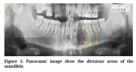
Each sample was divided according to the type radiographic technique into CBCT and OPG. The readings of each sample were achieved at different mandible regions; second premolar area (A), first molar area (B), second molar area (C) and distal to second molar area (D) as shown in Figure 1.

Figure 1. Panoramic image show the divisions areas of the mandible.
The samples were divided according to the gender into; male and female.
These areas were organized and divided by using dental software application and obtaining reconstructed panorama and cross sectional views to optimize the center of each examined view. The observer repeats the reading 3 times with one week interval between each reading.
The visibility of the MC was registered as either clear (C) or unclear (UC), in both CBCT and panoramic view, respectively, to differentiate the canal from surroundings, such as bone marrow spaces. The data of each examined area were clustered together so that each hemi-mandible received an overall visibility, then compared that assessed by CBCT with data that assessed by OPG for both gender (male and female).
The collected data was analyzed statistically by SPSS software program to assess the statistical significant differences between groups measured.
Results
The study results showed higher clear visibility percentage of the mandibular canal (260 hemimandible from 400) at region A with 65% by the CBCT than by OPG images (94 hemi-mandible) which was 23%. The region B showed 56% (224 hemi-mandible from 400) clear visibility by CBCT which was higher clear visibility than images by OPG which was 46% (184 hemi-mandibular from 400). The percentages at region C showed was 47% (188 hemi-mandible from 400) clear visibility by the CBCT which is less than OPG which was 51% (204 hemi-mandibular). The region E showed 42% (186 hemi-mandible from 400) clear visibility of the hemi-mandibles by CBCT while 73% (292 hemi-mandibular) by OPG Table 1 and Table 2.
| Type of x-ray | Regions | |||
|---|---|---|---|---|
| A | B | C | D | |
| OPG | 23% | 46% | 51% | 73% |
| CBCT | 65% | 56% | 47% | 42% |
Table 1: Percentage of hemi-mandibles clearance and MC visibility according to the regions by OPG and CBCT.
| Area | CBCT | OPG | P- value |
|---|---|---|---|
| A | 65% | 23.30% | P<0.05S |
| B | 56% | 46% | P<0.05S |
| C | 47.70% | 51% | P>0.05NS |
| D | 42% | 73% | P<0.05S |
Table 2: Multiple comparisons between CBCT and OPG of hemimandibles clearance and MC visibility percentages.
There were statistical differences between CPCT and OPG images at the region A, B and D while at the region C there was no significant difference as shown in Table 2.
When the CBCT results of males compared to those of females, the Followings data were observed, as showed in Table 3:
| area | Male | female | p-value |
|---|---|---|---|
| A | 65% | 63% | P > 0.05NS |
| B | 58% | 55% | P > 0.05NS |
| C | 50% | 51% | P > 0.05NS |
| D | 44% | 42% | P > 0.05NS |
Table 3: Percentage of hemi-mandibles clearance and visibility of mandibular canal according to the region and comparison between male and female by using CBCT.
✓ The visibility of the mandibular canal in area (A) was identified clearly 65% of male (130 hemi-mandible from 200), and 63% of female (126 hemi-mandibular from 200) which showed a non-significant difference between them.
✓ The visibility of the mandibular canal in area B was identified clearly in 58% (116 hemimandible from 200) in male, and 55% (110 hemi- mandibular from 200) in female, (The difference is statistically non-significant).
✓ The visibility of the mandibular canal in area C was identified clearly in 50% (100 hemimandible from 200) in male, and 51% (102 hemimandibular from 200) in female, (The difference is statistically non-significant).
✓ The visibility of the mandibular canal in D area was identified clearly in 44% (88 hemimandible from 200) in male, and 42% (84 hemimandibular from 200) in female (The difference is statistically non-significant).
Discussion
This study is the first one in Iraq that concerned to the mandibular canal visibility by CBCT in comparison with OPG. The MC is a significant anatomical structure and fundamental requirement for preoperative planning of surgical procedures involving the posterior mandible to minimize the possible complications during surgery.
In this study, detection of the MC course was assessed by using two types of imaging modalities; CBCT and panoramic radiograph. The results were presented the difference in the clearance of canal visibility throughout the normal anatomical landmarks. The new CBCT imaging technologies has allowed the investigation of anatomical structures in different plans without image overlapping or misdiagnosis with other structures (such as bone marrow). CBCT is a providing technique for the detailed diagnosis of bony anatomical land marks, with good resolution as compared with OPG and low radiation dose as compared with CT scan [7-14].
Oliveira-Santos et al in 2011 performed a study to assess the visibility of MC by CBCT, and reported that the visibility was increase with moving posteriorly (distal to mental foramen). The dis agreement with this study may be due to small sample size used by the authors or the different analytical method used.
Jung and Cho in 2014 found that visibility of MC by CBCT was better than by OPG and the visibility of canal was decreased by moving further posteriorly. This was disagreed with the results of this study and this may be due to differences evaluation technique or age of patients which may affect the visibility of canal, or reduced bone density and trabeculation.
The results of the present study showed lower percentage for MC visualization by OPG and this was agreed with Naitoh et al., Jung et al, Lindh et al. CBCT reported superior results compared to panoramic images for the identification of the mandibular canal, this agreed with Kamrun et al. in confirmed that the visibility of cross-sectional CT images was significantly higher than that of panoramic images of the mandibular canal.
In the present study, the mandibular canal was clearly visible on CBCT cross-sectional images of more anterior region than posterior region. Visibility decreased towards the posterior teeth. These findings are in disagreements with those previous study that found more clear mandibular canal posteriorly [15], this difference may be due to that previous studies did not use CBCT therefore they didn’t reach to clear radiographic visibility of the mandibular canal near the mental foramen due to the lack of definite walls in the anterior portion of the canal [16-18].
This study represent the importance of CBCT to clarify the anatomical land mark and mandibular canal with non - significant result between male and female. The CBCT images were more suitable images for the visualization of the mandibular canal, the identification of this structure seems to be more linked to the bone density of its walls, And more dependable on anatomic features of the canal itself than on the technique used9. Thus, the degree of difficulty should be expected in the visuilization of the canal on other imaging modalities [19-23].
In the study, the visualization of the mandibular canal on CBCT cross-sectional images was considered an easy procedure as compared with OPG especially in mental area, which indicates that importance of CBCT in visualize and diagnose the anatomical land mark of oral and maxillofacial region.
Conclusion
The mandibular canal visibility on CBCT crosssectional images is better than that in OPG. However, the visibility of the canal from its surrounds became less clear towards the posterior region of mandible. The results showed a high statistical significant differences between the two imaging technique with more clearance and visibility of the MC by CBCT. Differences in visibility of the mandibular on CBCT crosssectional images between male and female were statistically non-significant.
Recommendations
✓ Since the results of this study may not be generalized to the entire Nigerian and African population, we recommend the need to use the instrument to test for levels of stress and its sources at a multicenter level nationally and continentally.
✓ Further studies should be developed to compare the findings of the MSSQ-20 with other established instruments for stress evaluation.
✓ Findings from past, present, and future studies should be obtained and synchronized towards the formation of useful stress management strategies and guide for curricula review, policy draft and implementation, decision making, student support schemes, and guidance and counseling (G&C) services.
References
- Kim TS, Caruso JM, Christensen H, et al. A comparison of cone-beam computed tomography and direct measurement in the examination of the mandibular canal and adjacent structures. J Endodont 2010; 36:1191-1194.
- Akbulut N, Akbulut S, Oztas B, et al. Importance of bifid mandibular canal in implantology and in oral surgery: Review of the literature and report of three cases. Cumhuriyet Dent J 2017; 20:198-203.
- Libersa P, Savignat M, Tonnel A. Neurosensory disturbances of the inferior alveolar nerve: A retrospective study of complaints in a 10-year period. J Oral Maxillofac Surg 2007; 65:1486-1469.
- Angelopoulos C, Aghaloo T. Imaging technology in implant diagnosis. Dental Clinics. 2011; 55:141-158.
- Boeddinghaus R, Whyte A. Current concepts in maxillofacial imaging. Eur J Radiol 2008; 66:396-418.
- Koong B. Cone beam imaging: Is this the ultimate imaging modality?. Clin Oral Implants Res 2010; 21:1201-1208.
- Angelopoulous C, Thomas SL, Hechler S, et al. Comparison between digital panoramic radiography and cone-beam computed tomography for the identification of the mandibular canal as part of presurgical dental implant assessment. J Oral Maxillofac Surg 2008; 66:2130–2135.
- Kamburoğlu K, Kılıç C, Özen T, et al. Measurements of mandibular canal region obtained by cone-beam computed tomography: A cadaveric study. Oral Surg Oral Med Oral Pathol Oral Radiol Endodontol 2009; 107:e34-42.
- Liang X, Jacobs R, Hassan B, et al. A comparative evaluation of cone beam computed tomography (CBCT) and multi-slice CT (MSCT): Part I. On subjective image quality. Eur J Radiol 2010; 75:265-269.
- Lou L, Lagravere MO, Compton S, et al. Accuracy of measurements and reliability of landmark identification with computed tomography (CT) techniques in the maxillofacial area: a systematic review. Oral Surg Oral Med Oral Pathol Oral Radiol Endodont 2007; 104:402-411.
- Monsour PA, Dudhia R. Implant radiography and radiology. Australian Dent J 2008; 53:S11-25.
- Scarfe WC, Farman AG, Sukovic P. Clinical applications of cone-beam computed tomography in dental practice. J Canadian Dent Assoc 2006; 72:75.
- Tantanapornkul W, Okouchi K, Fujiwara Y, et al. A comparative study of cone-beam computed tomography and conventional panoramic radiography in assessing the topographic relationship between the mandibular canal and impacted third molars. Oral Surg Oral Med Oral Pathol Oral Radiol Endodontol 2007; 103:253-259.
- Ziegler CM, Woertche R, Brief J, et al. Clinical indications for digital volume tomography in oral and maxillofacial surgery. Dentomaxillofac Radiol 2002; 31:126–130.
- Oliveira-Santos C, Capelozza AL, Dezzoti MS, et al. Visibility of the mandibular canal on CBCT crosssectional images. J Applied Oral Sci 2011; 19:240-243.
- Denio D, Torabinejad M, Bakland LK. Anatomical relationship of the mandibular canal to its surrounding structures in mature mandibles. J Endodont 1992; 18:161-165.
- Gowgiel JM. The position and course of the mandibular canal. J Oral Implantol 1992; 18:383-385.
- Lindh C, Petersson A. Radiologic examination for location of the mandibular canal: A comparison between panoramic radiography and conventional tomography. Int J Oral Maxillofac Implants 1989; 4.
- Anderson LC, Kosinski TF, Mentag PJ. A review of the intraosseous course of the nerves of the mandible. J Oral Implantol 1991; 17:394-403.
- Naitoh M, Katsumata A, Kubota Y, et al. Relationship between cancellous bone density and mandibular canal depiction. Implant Dent 2009; 18:112-118.
- Wadu SG, Penhall B, Townsend GC. Morphological variability of the human inferior alveolar nerve. Clinical Anatomy 1997; 10:82-87.
- Jung YH, Cho BH. Radiographic evaluation of the course and visibility of the mandibular canal . Imaging Sci Dent 2014; 44:273–278.
- Kamrun N, Tetsumura A, Nomura Y, et al. Visualization of the superior and inferior borders of the mandibular canal: A comparative study using digital panoramic radiographs and cross-sectional computed tomography images. Oral Surg Oral Med Oral Pathol Oral Radiol 2013; 115:550-557.
Author Info
Resha Jameel1*, Areej Ahmed Najm1 and Firas Abdulameer Farhan2
1Department of Oral and Maxillofacial Radiology, Collage of Dentistry, University of Baghdad, Baghdad, Iraq2Department of prosthodontics, college of dentistry, University of Baghdad, Baghdad, Iraq
Citation: Resha Jameel, Areej Ahmed Najm, Firas Abdulameer Visibility of mandibular canal on CBCT cross-sectional images in comparison with panoramic radiograph (Retrospective study), J Res Med Dent Sci, 2021, 9 (2): 290-294.
Received: 04-Dec-2020 Accepted: 18-Jan-2021
