Research - (2021) Volume 9, Issue 2
Enhancement of Dental X-rays Images Using Image Processing Techniques
Yousif Mohamed Y Abdallah1*, Nouf H Abuhadi2 and Maryam Hasan Hugri3
*Correspondence: Yousif Mohamed Y Abdallah, Department of Radiological Science and Medical Imaging, College of Applied Medical Science, Majmaah University, Saudi Arabia, Email:
Abstract
Background: Medical imaging uses to clinical evaluation of the dental anatomy and pathology. Billions of images are performed globally. Imaging processing technique is used for helping accuracy of diagnosis by increasing the image quality. The dental images consider as a simple mixture of (unwanted) background information, diagnostic information, and noise. Aim: This paper is performed to enhance the dental images quality by denoising and detecting the dental structures. Materials and methods: This study design is experimental study done on dental images who are preliminarily diagnosed as having different oral diseases and is focused on using the image processing techniques to enhance the image quality of dental x-rays (OPG and Periapical) A convenience sampling was done and dental images were collected and then were treated by using MatLab program. Data analysis was done by using MatLab and statistical analysis was done by measuring precision and recall computation frame. Results: The quantitative analysis had done using both precision and recall computation. The results were 97.7 ± 8.16. This method presented capacities cumulative of recognition of the dental structures tissue with high precision rate and helps in enhancing the dental images.
Keywords
Dental x-rays, MatLab, Texture analysis
Introduction
The patient's intraoral teeth are relatively comfortable and acceptable overall radiation dose, but in some cases it is not optimally dental. However, it provides valuable information and far exceeds clinical outcomes, often promoting broader review strategies as the basis for treatment planning. Clear radiographs of the teeth and jaws, however, decreases the risk of incomplete, possibly incorrect examinations, leading to the worst possible misconduct. The X-ray spectrum always increases the horizon by increasing knowledge of radiation by dentists, thereby increasing their ability to identify normal and pathological conditions. Besides better understanding the connections between systemic and oral medical conditions [1-4], new treatment planning approaches could be opened. The radiation doses used or transferred to an auxiliary dentist are responsible for the quality of the x-rays obtained. The responsible person should ensure that they are properly trained and legally certified when assigned to the job [5,6]. To protect patients and staff from radiation over-exposure, dental indications and procedures must be understood. They should not only be high-quality x-ray manufacturers. Each patient should receive the maximum level of X-ray radiation and the lower dose of X-ray dental to ensure that all training standards are maintained. Crucial here. Picture that can be applied as image, painting, or sketch to a physical image. But a physical rather than mental idea or concept can also be applied. When asked to imagine an object, the apple comes to mind [7-9]. It enables apple concept to be understood. Obviously, an apple's photographic visual appearance is only one aspect. Apple's taste, smell, or feeling that we remember is not represented by our experience [8-10].
The image spans two elements. The amount of data that can be transmitted is limited. Especially the sound effect can lose details of the fine structure. Noise can see a term unlikely to be used in a silent visual image. Because of this term, radio technology is likely to damage the receiving quality of radio signals in low background noise (shoots and whistles) and the receiving signal amplification. Such RDI effects are very similar to image information noise. Under optimal conditions, the signal magnitude is significantly higher than the noise. High signal/noise [11-14]. Signal/noise ratio is legal and much information is lost in adverse circumstances. Disruption of an x-ray image is unlikely due to accidental x-ray chamber exposure or poor storage. The image seems fog or fog. The image data content decreases. Some information is lost, but it's hard to see what's left. Also, fogging doesn't supply X-ray data. Such fog is a picture noise example [15-17]. A dental images show several noises.. There are many small, overlaid white spindles on the screen. This leads to another decline in photo data content [4,7,18-20]. For example, an electronic system may appear due to lower design leading to electronic noise. However it is important to note that the signals are often too weak and the puzzle doesn't lack space. In this case, the image appears to have many small black spots in an X-ray image, commonly called quantic mottle or quantic noise. The image feature must appear different from its environment in this case. When X-rayed adjacent structures have different optical densities (gray shade). The image structure must be different (brightness) [4,7]. The term contrast describes such differences; the structure is hard to understand and is called high (good). The framework cannot recognize small differences [9,10].
Materials and Methods
The digital scanner used MatLab Image Processing to evaluate panoramic image improvements and contrasts. The scanned image is saved for TIFF file format image quality. Gray concentration data was used to improve contrast between soft tissue, which can be distributed linearly or not by gray frequency and sound from original images. Figures 2-1 shows the block diagram in the proposed paper. This research uses preprocessing technology stepby- step X-ray images. This diagram explains the workflow (Figure 1). Hospitals and RGB transformation. Two filtration algorithms filter panoramic photos. Anisotropic filtering for filtering algorithms. Results compared with PSNR and MSE values. Image enhancement filtering. The image improves contrast, reduces noise, and sharpens the performance edge. Improved contrast, detail sharpening, and visibility are goals for image enhancement. Some equalization algorithms exist (CLAHE). The researchers used the algorithm for limited histogram equalization. For X-ray output, PSNR offers the best PSNR value (CLAHE).
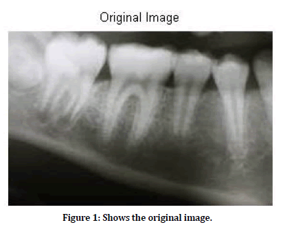
Figure 1. Shows the original image.
Results
This study provides a new approach for separate panoramic image noise processing to reduce redundancy of image data (MatLab version R2020b). In addition to emphasizing the role of the proposed approach (noise variations), the importance of using radiological imaging technologies is emphasized in maintaining the overall image appearance, maintaining the diagnostic level, and detecting small and small contrast data for the image's diagnostic content. Figure 1 shows the original dental images before to be processed by image processing program. Gray conversion Figure 2 original RGP image. Figure 2 Shows processed gray scale image. Those images were processed by flatted-field correction to reduce the PNSR in the radiographs. This gray color image applied into anisotropic filtering method the output is in Figure 2 and also applied in median filtering algorithms the output is in Figures 3A-3D). Figures 4A-4D Shows histogram of the median filtered images. Figure 5 shows edge detection using conventional methods. Figure 6A shows edge detection using simplest syntax. Figure 6B Shows edge detection using block borders.
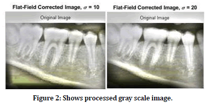
Figure 2. Shows processed gray scale image.
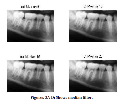
Figures 3A-D: Shows median filter.
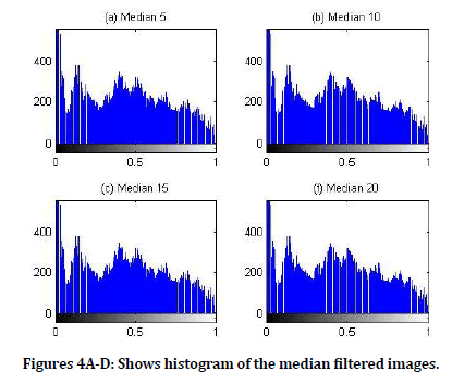
Figures 4A-D: Shows histogram of the median filtered images.
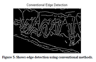
Figure 5. Shows edge detection using conventional methods.
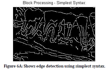
Figure 6a. Shows edge detection using simplest syntax.
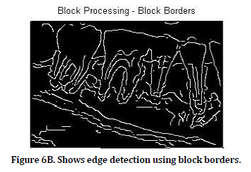
Figure 6b. Shows edge detection using block borders.
Discussion
In real X-ray images, sound content is substantially consistent. Two panoramic acquisitions identify the problem; one (Figure 1) is a technique for dental scanning, while another is an X-ray imaging technique (figure 2). Figure 2 shows the ideal view to enhance and remove panoramic x-rays from the mixture. Preprocessing aims to improve image quality. They should be filtered and detected if too boring or absurd. Filters eliminate a large image such as rendering or image scale: improving or detecting image limits. However, pictures are usually poor due to different variables. Researchers use preprocessing images like noise and bubbles to eliminate artifacts and degradation. Several non-linear smoothing filters. Although their properties and application areas were studied in detail, Fourier cannot usually submit them for analysis. Scientists used medium and anisotropic filters. The research system used medium and anisotropic filter algorithms. A filter that respects image edges. The median filter resembles the average noise reduction filter (it is a simple, intuitive and easy to implement method of smoothing images, i.e. reducing the amount of intensity variation between one pixel and the next. It is often used to reduce noise in images). As the average filter, the media filter reduces image noise. Often however, useful image data are better maintained than average filter. Each pixel is seen as the average filter and looks around its neighbors to see if they represent their surroundings. The average pixel, not the adjacent average, is replaced. The media is calculated first when all pixels are sorted numerically, replacing the center pixel. In 10 different X-ray images, anisotropic and medium filtering algorithms are used to calculate MSE and PSNR.
Technical edge detection
As large images are processed, standard image processing strategies can break down. Images can be too large to load memory or memory. The following problems can be managed slowly for large images: from one region to another on the computer, read, process and record results. A block procedure facilitates this. Enter the block's image, block size and process. Blockproc divides the image into one block and processes it. Blockproc returns the new disk fire or Figure 5 to your memory.
Each Y-array element is removed by Z = subtracted(X,Y) and the respective differences of the resulting Z-screen. X and Y are real arrays of the same size and class, unsaved, and doublescale. When X is wrong, the RO is category X, doubling in this case. If X is an integer array, X reduces outputs that are larger than an integer range and extracts the fractional numbers.
Conclusion
The experimental study proposed a new approach for independent, identical dental noise distribution and reduced image data redundancy using imaging techniques (MatLab version R2009a). Not only the role of the proposal approach, but also the diagnostic content and the role of radiological use of imaging technology showed the overall appearance of the image with its diagnostic content and small and low contrast details (noise variation). This has caused the edges to be identified near the initial storage output. Other artifacts were on the border. Canny Edge Detector uses various padding techniques. This paper concludes that an attempt to understand the issue supports a new approach to viewing and filtering panoramic images as a combination of (unwanted) background information, diagnostic data and information about noise. Only along the image boundary supports zero padding with the Separating Blind Sources, Border Detection, and Images. Noise detection is a complex and hardto- detect process for powerful image analysis with the naked eye.
References
- Lin PL, Lai YH, Huang PW. An effective classification and numbering system for dental bitewing radiographs using teeth region and contour information. Pattern Recognition 2010; 43:1380–1392.
- Jain AK, Chen H. Matching of dental X-ray images for human identification. Pattern Recogn 2004; 37:1519–1532.
- Banumathi A, Vijayakumari B, Raju S. Performance analysis of various techniques applied in human identification using dental X-rays. J Med Syst 2007; 31:210-218.
- Said EH, Diaa EM, Nassar GF, et al. Teeth segmentation in digitized dental X-ray films using mathematical morphology. Transactions Information Forensics Security 2006; 1:178–189.
- Nomir O, Abdel-Mottaleb M. Fusion of matching algorithms for human identification using dental X-ray radiographs. IEEE Transactions on Information Forensics and Security 2008; 3:223-233.
- Nomir O, Abdel-Mottaleb M. Human identification from dental X-ray images based on the shape and appearance of the teeth. IEEE Transactions Information Forensics Security 2007; 2:188-197.
- Nomir O, Abdel-Mottaleb M. Hierarchical contour matching for dental X-ray radiographs. Pattern Recognition 2008; 41:130-138.
- Fahmy G, Nassar D, Haj-Said E, et al. Towards an automated dental identification system (ADIS). InInternational conference on biometric authentication. Springer, Berlin, Heidelberg 2004; 789-796.
- Hosntalab M, Zoroofi RA, Tehrani-Fard AA, et al. Automated dental recognition in MSCT images for human identification. Fifth international conference on intelligent information hiding and multimedia signal processing IEEE 2009.
- Aeini F, Mahmoudi F. Classification and numbering of posterior teeth in bitewing dental images. 3rd International conference on advanced computer theory and engineering (ICACTE) 2010.
- Phong-Dinh V, Bac-Hoai L. Dental radiographs segmentation based on tooth anatomy. IEEE International conference on research, innovation and vision for the future in computing & communication technologies university of science—Vietnam National University, Ho Chi Minh City 2008.
- Shah S, Abaza A, Ross A, et al. Automatic tooth segmentation using active contour without edges. IEEE, Biometrics symposium, 2006
- Hofer M, Marana AN. Dental biometrics: Human identification based on dental work information. XX Brazilian symposium on computer graphics and image processing IEEE 2007; 1530–1834/07.
- Lifeng He, Yuyan Chao, Kenji Suzuki. An efficient first-scan method for label-equivalence-based labeling algorithms. Pattern Recogn Lett 2010; 31:28–35.
- Dawoud NN, Samir BB, Janier J. Fast template matching method based optimized sum of absolute difference algorithm for face localization. Int J Comp Appl 201; 18.
- Oprea S, Marinescu C, Lita I, et al. Image processing techniques used for dental x-ray image analysis. In2 008 31st international spring seminar on electronics technology IEEE 2008; 125-129.
- Olsen GF, Brilliant SS, Primeaux D, et al. An image-processing enabled dental caries detection system. In 2009 ICME international conference on complex medical engineering 2009; 1-8.
- Kiattisin S, Leelasantitham A, Chamnongthai K, et al. A match of x-ray teeth films using image processing based on special features of teeth. In 2008 SICE Annual Conference 2008; 35-39.
- https://www.wiley.com/en-in/Dental+Caries%3A+The+Disease+and+its+Clinical+Management%2C+3rd+Edition-p-9781118935828
- Paul Fotek. Florida Institute for Periodontics & Dental Implants. Published in Medical Plus, A service of the U.S. National Library of Medicine National Institutes of Health.
Author Info
Yousif Mohamed Y Abdallah1*, Nouf H Abuhadi2 and Maryam Hasan Hugri3
1Department of Radiological Science and Medical Imaging, College of Applied Medical Science, Majmaah University, 11952, Majmaah, Saudi Arabia2Department of Diagnostic Radiology, College of Applied Medical Sciences, Jazan University, Jazan, Saudi Arabia
3Department of Maxilloofacial Surgery and Diagnostic Sciences, College of Dentisry, Jazan University, Jazan, Saudi Arabia
Citation: Yousif Mohamed Y Abdallah, Nouf H Abuhadi, Maryam Hasan Hugri, Enhancement of Dental X-rays Images Using Image Processing Techniques, J Res Med Dent Sci, 2021, 9 (2): 12-16.
Received: 15-Dec-2020 Accepted: 18-Jan-2021
