Case Report - (2025) Volume 13, Issue 3
Florid Cemento Osseous Dysplasia: A Case Report
*Correspondence: Reema A Sharaf, Department of Restorative Dentistry, Prince Sultan Medical Military City, Riyadh, Saudi Arabia, Email:
Abstract
Florid Cemento-Osseous Dysplasia (FCOD) is a fibro-osseous disease characterised by the replacement of normal bone with poorly cellularized cementum-like materials and cellular fibrous connective tissues through a reactive process. The condition is limited to the places where teeth are present or absent, and it often affects both sides with symmetrical involvement. This case report discusses an unusual occurrence of a 21-year-old female who had many impacted permanent teeth along with multiple radiopaque lesions in the periapical area. The Orthopantomogram (OPG) revealed radiopaque lesions in the locations where teeth were missing. FCOD is radiographically identified by the presence of numerous masses composed of a combination of radiopaque components. There could be a circular area of decreased density on X-rays, mainly at the tips of healthy teeth. As the lesions develop, they typically grow denser on X-rays. The management of dental caries and removal of teeth that are beyond salvage have been successfully carried out. The patient was receiving routine clinical and radiological monitoring. When a radiolucent or mixed lesion appears at the periapical area of a tooth with a live pulp, we recommend using panoramic radiography to distinguish between an inflammatory periapical lesion and a lesion of Cemento-Osseous Dysplasia (COD).
Keywords
Florid Cemento-Osseous Dysplasia (FCOD), Radiopaque lesions, Orthopantomogram (OPG), Panoramic radiography, X-rays
Introduction
Fibro-osseous lesions of the jaws encompass fibrous dysplasia, ossifying fibroma, and Cemento-Osseous Dysplasia (COD). Cemento-Ossifying Dysplasia (COD) typically manifests in the alveolar regions of the maxilla and mandible and is likely the prevailing fibro-osseous abnormality found in clinical settings [1]. There exist three distinct categories of COD lesions, specifically focal, periapical, and Florid COD (FCOD). Treatment is typically unnecessary for the COD lesion, but surgical recontouring or complete excision is necessary for the other two fibro-osseous lesions. Hence, it is crucial to accurately determine the differential diagnosis of three fibro-osseous lesions, since an incorrect diagnosis could result in unwarranted endodontic therapy, incisional biopsy, or surgical excision. This article presents a case of Florid Cemento-Osseous Dysplasia (FCOD) in a 21-year-old female patient who sought treatment at an emergency dentistry clinic because to recurring facial swelling and pain in her upper tooth. The patient was completely uninformed of her condition. The case was initially identified by a clinical examination, which was followed using Orthopantomogram (OPG) for additional investigation. We will analyze the diagnosis and explore the potential therapy alternatives for this specific case.
Case Presentation
A 21-year-old Saudi woman presented herself to the emergency department of a dentistry centre. She presented with pain and recurring swelling in the upper right side of her face for a duration of one month. The patient had a past record of repeated facial swellings in connection with her upper right molar area. There were no significant contributions from the family and medical history.
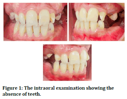
The extraoral examination identified facial asymmetry and edoema in the upper region of the face, without any noticeable changes in colour or warmth. The intraoral examination revealed the absence of teeth 18, 17, 15, 14, 13, 23, 25, 26, 27, 35, 34, 33, 44, and 45 (Figure 1).

Figure 1: The intraoral examination showing the absence of teeth.
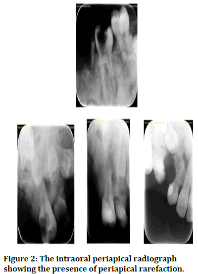
The teeth with caries are numbered as follows: 16, 28, 36, 37, and 47. The primary teeth that were kept are numbered 53, 55, and 73, while the surviving root of primary tooth 63 was also retained. The individual had inadequate oral hygiene, resulting in the buildup of plaque. Tooth 16 exhibited caries and was non-vital. The intraoral periapical radiograph revealed the presence of periapical rarefaction (Figure 2).

Figure 2: The intraoral periapical radiograph showing the presence of periapical rarefaction.
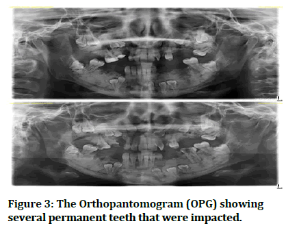
Obtaining periapical radiographs for the remaining teeth posed challenges due to the constricted maxillary arch and the patient's low tolerance. The Orthopantomogram (OPG) revealed several permanent teeth that were impacted, as well as extensive areas of radio opacity in both the upper and lower dental arches, namely in the periapical regions of all teeth in both quadrants of the upper and lower arches (Figure 3).

Figure 3: The Orthopantomogram (OPG) showing several permanent teeth that were impacted.
Radio opaque rims are often found in connection with impacted teeth. The impacted teeth were profoundly positioned, extending to the lower boundary of the mandible in the lower jaw and reaching the maxillary sinus in the upper jaw. The patient exhibited a complete lack of awareness of her dental status, and none of her dental visits included any information or communication on the state of her teeth. Tooth 16 underwent endodontic treatment, and the patient remains symptom-free to this day. The diagnosis of FCOD with a chronic periapical infection was established based on clinical and radiological evidence. The patient has been undergoing regular follow-up for the past six months, and there have been no changes observed in the size, number, or behaviour of the lesions. We refrained from doing a biopsy due to the patient's stable condition.
Discussion
The name Florid Cemento-Osseous Dysplasia (FCOD) was initially proposed by Melrose, et al. in 1976 to characterize a disorder that exhibits abundant, multi-quadrant masses of cementum and/or bone in both jaws, and occasionally, simple bone cavity-like lesions in the affected quadrant [2]. The term 'florid' was coined to reflect the vast and widespread signs of the disease in the jaws. FCOD does not have any additional abnormalities outside of the jaw and there are no abnormalities in the blood chemistry of patients [3]. The disease exhibits a notable inclination towards occurring on both sides, frequently manifesting symmetrically in the jaws. If the lesions are of significant size, there may be an observable enlargement of the jaw, along with indications of persistent pain or discharge in the affected region [4].
In the case of an asymptomatic patient, it is advisable to closely monitor the patient without resorting to surgical intervention, as there are diagnostic measures available [4,5]. The purpose of sharing this intriguing example is to demonstrate the deviation from the typical radiography presentation. FCOD lesions are often discovered incidentally in most cases. Therefore, an oral physician must possess a thorough understanding of the potential occurrence of cortical bone growth or localized infection with drainage.
The procedure of differential diagnosis is particularly important when there are coincidental abnormalities such as odontogenic infections or chronic diffuse osteomyelitis, neoplasia, and bone dysplasia, which can all lead to comparable radiographic alterations in the mandible. Therefore, it is imperative that we carefully evaluate all the diagnostic information at our disposal, particularly the radiographic characteristics that play a crucial role in making the diagnosis [6]. In addition, FCOD can render the jaws vulnerable to osteomyelitis and pathological fracture. These sequelae increase the importance of ongoing follow-up, making it obligatory.
Conclusion
To summarize, this case report emphasizes the clinical and radiological characteristics of FCOD, an uncommon and noncancerous fibro-osseous abnormality that mostly impacts the jawbones. FCOD commonly manifests as several dense masses with a distinctive "cotton-wool" look on X-ray images. These masses are frequently found by chance during regular dental check-ups. While FCOD is typically without symptoms, it can sometimes result in problems such as infection and displacement of teeth. Clinicians must possess knowledge of FCOD and its unique radiological and clinical characteristics to ensure accurate diagnosis and effective treatment. Moreover, it is crucial to provide patients with education and reassurance, as FCOD is a non-malignant illness that does not necessitate active management unless symptoms or consequences arise.
Additional investigation is necessary to gain a deeper comprehension of the causes and development of FCOD, as well as to define definitive criteria for its diagnosis and treatment. with expanding our understanding of this condition, we may better the quality of care provided to patients, improve the results of treatment, and reduce unnecessary treatments, so ensuring the best possible oral health and well-being for individuals affected with FCOD.
References
- Neville BW, Damm DD, Allen CM, et al. Oral and maxillofacial pathology. 3rd Ed. Philadelphia: Saunders. 2009; 613–677, 701–740.
- Brown KE. Fabrication of ear prosthesis. J Prosthet Dent 1969; 21:670–676.
[Crossref] [Google Scholar] [PubMed]
- Melrose RJ, Abrams AM, Mills BG. Florid osseous dysplasia. Oral Surg 1976; 41:62–82.
[Crossref] [Google Scholar] [PubMed]
- Waldron CA. Fibro-osseous lesions of the jaws. J Oral Maxillofac Surg 1985; 43:249-262.
[Crossref] [Google Scholar] [PubMed]
- Waldron CA. Fibro-osseous lesions of the jaws. J Oral Maxillofac Surg 1993; 51:828-835.
- Jeres W, Banu B, Winson B, et al. Florid cemento-osseous dysplasia in young Indian women: A case report. Brit Dent J 2005; 198:477-478.
[Crossref] [Google Scholar] [PubMed]
Author Info
Department of Restorative Dentistry, Prince Sultan Medical Military City, Riyadh, Saudi ArabiaCitation: Reema A Sharaf. Florid Cemento Osseous Dysplasia: A Case Report, J Res Med Dent Sci, 2024, 12 (01): 038-040.
Received: 19-Jan-2024, Manuscript No. JRMDS-24-128082; , Pre QC No. JRMDS-24-128082 (PQ); Editor assigned: 22-Jan-2024, Pre QC No. JRMDS-24-128082 (PQ); Reviewed: 29-Jan-2024, QC No. JRMDS-24-128082; Revised: 12-Feb-2024, Manuscript No. JRMDS-24-128082 (R); Published: 23-Feb-2024
