Research - (2022) Volume 10, Issue 1
Histological Evaluation of the Effect of Local Application of Bone Salioprotein on Bone Healing on Rat
Mayada Kamel Jaafar* and Enas Fadhil Kadhim
*Correspondence: Mayada Kamel Jaafar, Department of Oral Diagnosis, College of Dentistry, University of Baghdad, Iraq, Email:
Abstract
Background: Bone may be basically exceedingly vascular, living, continually evolving mineralized connective tissue. It will be amazing to its hardness, flexibility furthermore regenerative capacity, as well as its characteristic growth mechanisms. Objectives: histological study to substantiate that bone salioprotein when applied locally on generated bone defect healing in rat tibia, it was very effectiveness. Material and method: In this study forty-eight albino male rats, weighting (300-400) gram, aged (6-8) months, will make utilized under control states for temperature, drinking and food utilization. The animals will subject for an intra-bony defect in medial side of tibiae bone, in control group (12 rats) the bone defect dealt with absorbable haemostatic sponge, same time the experimental group (12 rats) will be dealt with local application of 30 μl bone salioprotein settled toward absorbable haemostatic sponge. The rats were sacrificed during 7, 14, also 28 days after surgery (four rats to every time for each group). Results: Histological discoveries shown that bone defect dealt with local application of bone salioprptein demonstrates a promptly osteoid tissue formation with high cell number for osteoblast, osteocyte furthermore osteoclast. Conclusion: Those goals that bone salioprotein improve bone healing and increase osteogenic capacity; this proof done our information need aid pledging for could reasonably be expected clinical management done future.
Keywords
Bone, Bone salioprotein, Bone healing
Introduction
Bone may be a progressive tissue, which undergoes consistent remodelling all around life time and show regeneration properties after damage remodelling will be the result of the complex transaction between osteoclasts.
Furthermore osteoblasts, which secure bone resorption furthermore bone formation respectively, and will be controlled eventually by the coordinated action of different biochemical and biophysical stimuli [1].
Bone may be comprised by dry weight for around 28% kind I collagen and 5% non-collagenous matrix proteins for example, bone sialoprotein, osteocalcin, osteonectin, osteopontin, what’s more proteoglycans; Growth factor and serum proteins also are found to bone.
This organic matrix will penetrate toward substituted hydroxyapatite (Ca10[PO4]6[OH]2), which makes it dependent upon the remaining 67% of bone [2].
BSP is a significant non-collagenous protein in mineralizing connective tissues, for example, such that dentin, cementum, bone, and calcified cartilage tissues [3].
BSP will be generated all by osteoblasts, osteoclasts, osteocytes, and furthermore hypertrophic chondrocytes throughout bone morphogenesis [4].
Bone sialoprotein (BSP) will be a standout amongst the real extracellular matrix (ECM) proteins of the bone. Also belongs to the little integrin-binding ligand N-linked glycoprotein (SIBLING) group [5].
Bone sialoprotein might regulate cell tasks like proliferation, adhesion, apoptosis, angiogenesis, migration, and ECM remodeling [6].
“Absorbable Haemostatic Gelatin Sponges” would sterilize, absorbable gelatin porcine sponges utilized to hemostasis, by applying to a bleeding site furthermore not connected over defiled body regions.
They would off-white color and spongy in look. Those hemostasis those dying over (2–4) minutes, Liquefies to (2-5) days to contact for mucosa, Also it might have been absorbing in any event 35 times its identity or weight for liquids also blood [7].
Materials and Methods
Every one experimental process was conveyed out in understanding with the moral standards about dentistry college of Baghdad. In this study forty - eight albino male rats, weighting (300-400) gram, aged (6-8) months, will make utilized under control states of temperature, drinking and food stuff utilization.
The animals subjected for an intra-bony defect in medial side of tibiae bone , in control group (12 rats) the bone defect dealt with absorbable hemostatic sponge , same time the experimental group(12 rats) will be dealt with local application of 30 μl bone salioprotein settled by absorbable hemostatic sponge .
The rats were sacrificed at 7, 14, and 28 days after surgery (four rats to each time done each group).
In the research, the materials connected were:
Bone sialoprotein (Recombinant Mouse Bone Sialoprotein.
IBSP Protein (HisTag) Elabscience Company.
Surgical procedure
The animals were subjected to a surgical operation. The surgery was performed under a sterilize state What's more delicate method. Each animal might have been weighted on ascertain those dosage for general anesthesia that might have been provided for to it.
That general anesthesia might have been prompted by Intramuscular injection about xylazine 2% (0. 4 mg/kg B. W.), Also ketamine Hcl 50mg (40 mg/kg- B. W.) Additionally an anti-microbial cover for oxytetracycline 20% (0. 7 ml/kg) intramuscular injection might have been provided for.
Tibiae will make shaved also the skin will a chance to be cleaned with a mixture of ethanol and iodine. At that point a bit about cotton is damped for spirits. Incision made on the lateral side to expose medial side of the tibia, the skin furthermore fascia flap was reflected.
By instrument flying drilling, what’s more constant cooling for irrigated saline, a hole about 1.8mm diameter settled on with little round bur during a rotating speed of 1500 rpm with depth to the bone marrow.
Furthermore, those holes preparation; the operation site might have been washed for the saline result to uproot trash starting with the penetrating site. The preoperative and postoperative hurt supervision was done by organization of meloxicam 0.2 mg/kg body weight intramuscularly.
Histological specimen preparation
Those specimens were settled around 24 hours on 10% formalin. At that point set in result about formic corrosive should decalcify those specimens, furthermore might have been dehydrating eventually what's more embedded previously, wax (paraffin).
A segment of 4- 5μm might have been transformed in the regular architecture; with hematoxylin what's more eosin might have been stained. The light magnifying lens might have been utilized to assess histological examine.
Statistical analysis
For bone cells number in histological research calculate by Min., Mean, Max., F-test, S.D, and P-value.
Results
Histological findings
For control group: During 7-days duration, indicates inflammatory cells, deposition of osteoid tissue (Figure 1 and 2).
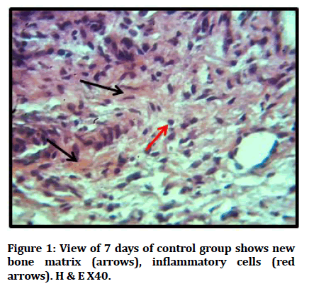
Figure 1: View of 7 days of control group shows new bone matrix (arrows), inflammatory cells (red arrows). H & E X40.
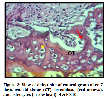
Figure 2: View of defect site of control group after 7 days, osteoid tissue (OT), osteoblasts (red arrows), and osteocytes (arrow head). H & E X40.
During 14 days duration, new immature bone trabeculae filled large number of irregularly arranged osteocytes and osteoblasts seen at peripheries of trabeculae. Reversal line separating old and new bone (Figures 3 and 4).
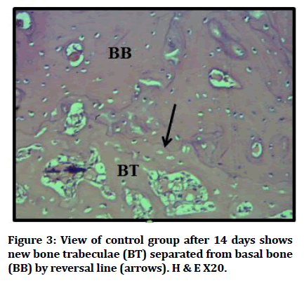
Figure 3:View of control group after 14 days shows new bone trabeculae (BT) separated from basal bone (BB) by reversal line (arrows). H & E X20.
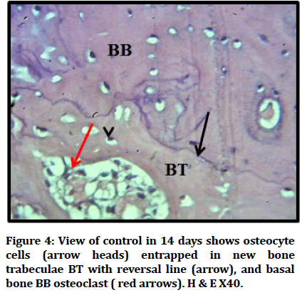
Figure 4:View of control in 14 days shows osteocyte cells (arrow heads) entrapped in new bone trabeculae BT with reversal line (arrow), and basal bone BB osteoclast ( red arrows). H & E X40.
During 28 days duration, mature bone almost filling the operating site, osteoblasts osteocytes, reversal line demarcating old and new bone (Figures 5 and 6).
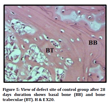
Figure 5:View of defect site of control group after 28 days duration shows basal bone (BB) and bone trabeculae (BT). H & E X20.
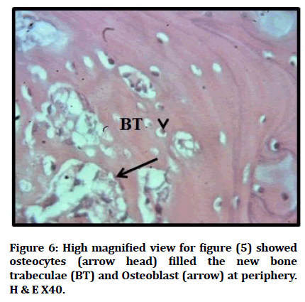
Figure 6:High magnified view for figure (5) showed osteocytes (arrow head) filled the new bone trabeculae (BT) and Osteoblast (arrow) at periphery. H & E X40.
For experimental group: During 7 day duration, shows deposition of bone matrix and trabeculated bone coalesce with cutting bone with reticular tissue, reticular cell within defect area rimmed by formative osteoblasts , and enclosing large number of osteocytes (Figures 7 and 8).
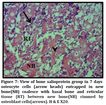
Figure 7:View of bone salioprotein group in 7 days osteocyte cells (arrow heads) entrapped in new bone(NB) coalesce with basal bone and reticular tissue (RT) between new bone(NB) rimmed by osteoblast cells(arrows). H & E X20.
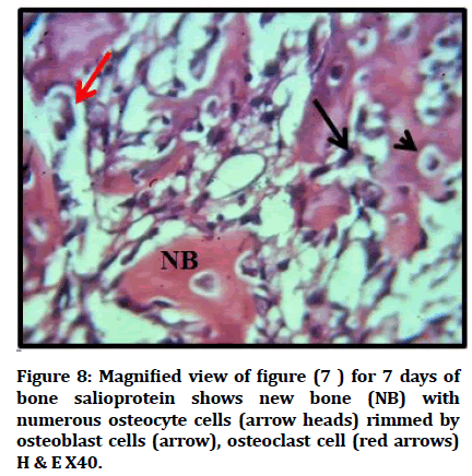
Figure 8:Magnified view of figure (7 ) for 7 days of bone salioprotein shows new bone (NB) with numerous osteocyte cells (arrow heads) rimmed by osteoblast cells (arrow), osteoclast cell (red arrows) H & E X40.
During 14 days duration, newly formed bone trabeculae enclosing areas of bone marrow, and including numerous regularly arranged osteocytes, osteoblasts seen at peripheries, osteoclast and reversal line also seen (Figures 9 and 10).
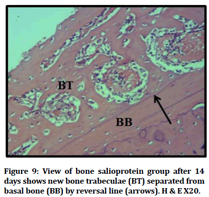
Figure 9:View of bone salioprotein group after 14 days shows new bone trabeculae (BT) separated from basal bone (BB) by reversal line (arrows). H & E X20.
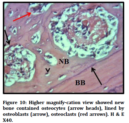
Figure 10:Higher magnify-cation view showed new bone contained osteocytes (arrow heads), lined by osteoblasts (arrow), osteoclasts (red arrows). H & E X40.
During 28 days duration, dense bone in defect site, osteocytes arranged in regular pattern around harversian canal, Osteoblasts seen at borders of bone (Figures 11 and 12).
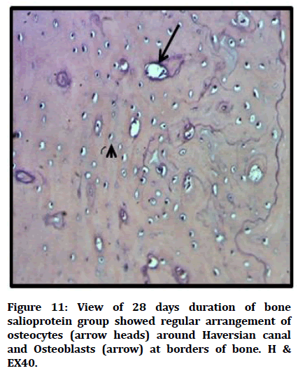
Figure 11:View of 28 days duration of bone salioprotein group showed regular arrangement of osteocytes (arrow heads) around Haversian canal and Osteoblasts (arrow) at borders of bone. H & EX40.
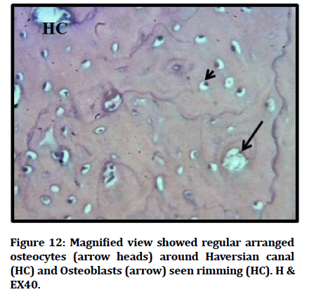
Figure 12:Magnified view showed regular arranged osteocytes (arrow heads) around Haversian canal (HC) and Osteoblasts (arrow) seen rimming (HC). H & EX40.
Analysis of statistical data
Histological analysis
Statistical analysis to bone cells mean number contain for osteoclast osteoblast, osteocyte for three 7, 14 Furthermore 28 interval, exhibits the bone salioprotein groups demonstrate an expanding mean quality with profoundly critical contrasts at compared with control groups (Table 1).
| Duration | Cells | Groups | Descriptive Statistics | Comparison | ||||
|---|---|---|---|---|---|---|---|---|
| Mean | S.D. | Min. | Max. | F-test | p-value | |||
| 7 days | OB | Cont | 34.825 | 12.012 | 28.25 | 52.8 | 1.544 | 0.254 |
| B | 42.623 | 3.319 | 39.22 | 45.5 | ||||
| OC | Cont | 36.05 | 3.007 | 33 | 40.2 | 5.715 | 0.011 | |
| B | 41.04 | 1.312 | 39.66 | 42.75 | ||||
| OCL | Cont | 0.375 | 0.206 | 0.2 | 0.6 | 0.416 | 0.745 | |
| B | 0.475 | 0.236 | 0.3 | 0.8 | ||||
| 14 days | OB | Cont | 30.708 | 3.48 | 27 | 34 | 16.739 | 0 |
| B | 21 | 1.414 | 19 | 22 | ||||
| OC | Cont | 33.83 | 5.97 | 28.66 | 39 | 6.053 | 0.009 | |
| B | 25.55 | 0.058 | 25.5 | 25.6 | ||||
| OCL | Cont | 0.8 | 0.216 | 0.5 | 1 | 12.466 | 0.001 | |
| B | 1.8 | 0.231 | 1.6 | 2 | ||||
| 28 days | OB | Cont | 17.25 | 3.202 | 14 | 20 | 1.365 | 0.3 |
| B | 12.25 | 2.723 | 8.5 | 15 | ||||
| OC | Cont | 30.33 | 1.359 | 29 | 31.66 | 15.053 | 0 | |
| B | 21.563 | 3.294 | 18.5 | 26 | ||||
| OCL | Cont | 0.3 | 0.346 | 0 | 0.6 | 0.227 | 0.875 | |
| B | 0.35 | 0.191 | 0.1 | 0.5 | ||||
Table 1: Descriptive statistics of the bone cells count (H&E) and groups’ difference in each duration.
Discussion
In introduce study was assessed the powerful of bone salioprotein applicated mainly which may be inspired once tentatively bone tibial harm to rats. The rat might have been utilized to a huge extent 38% t0 40% from animal fracture investigations to noticeable orthopaedic researches again the most recent decade [8].
That histological examination from bone areas indicated confirmation about osteoid bone in all planned groups after one week and immature bone spicules with osteoblasts seen at periphery more obviously in investigational group in agreement with discoveries about[(9] who specified that significant osteoid matrix synthesis and mineralization accelerated might have been toward early cell differentiation.
Microscopical finding demonstrated new bone formation by presence of numerous bone trabeculae and increase in osteoblasts number as contrasted with a control group. This discovering come to an agreement with Xu, [10] who showed that BSP might have been equipped to upgrade osteoblastic differentiation also new bone formation in vivo by stimulating the early bone mineralization.
Also Kruger et al. [11] found that in first week afterward surgery in a rat calvarial defect model. BSP–collagen scaffolds invigorated osteoblast differentiation, bone growth, and vascularization in vivo
Histological discoveies of the available study at 14 days uncovered new irregular bone trabeculae formation for both experimental and control groups at for distinction rate of bone deposition furthermore remodeling. Lacunae containing osteocytes were scattered within the newly formed bone trabeculae. This bring about concurrence for Monfoulet, et al. [12] who made drill-hole defects in the cortex about mouse femur What's more discovered the cortical hole might have been bridged for trabecularlike bone inside 2 weeks and no mineralized tissue might have been watched in the marrow.
Choi, et al. [13] investigated collagen-binding (CB) peptides determined starting with BSP were connected will a hydroxyl apatite scaffold, bone development might have been altogether invigorated to an in vivo rabbit calvarial defect inside two weeks.
In 28 days more ranges of bone deposition were watched with slim furthermore little bone trabeculae as accounted for by Amera, [14] same time those available outcomes demonstrated thick full grown bone in experimental group at 4weeks. This outcome assention with finding of Klein, (2020) [15] who investigated that an inclination towards an expanded bone formation in the BSP at 4 weeks then afterward scaffold implantation the examination of the control.
References
- Boyce BF, Li J, Xing L, et al. Bone remodeling and the role of TRAF3 in osteoclastic bone resorption. Front Immunol 2018; 9:2263.
- Nanci A. Ten cate's oral histology-e-book: development, structure, and function. Elsevier Health Sciences 2017.
- Maeda H, Wada N, Tomokiyo A, et al. Prospective potency of TGF-Ã?1 on maintenance and regeneration of periodontal tissue. Int Rev Cell Mol Biol 2013; 304:283-367.
- Maaroufi A, Khadem-Ansari MH, Khalkhali HR, et al. Serum levels of bone sialoprotein, osteopontin, and Ã?2-microglobulin in stage I of multiple myeloma. J Cancer Res Ther 2020; 16:98.
- Xu L, Zhang Z, Sun X, et al. Glycosylation status of bone sialoprotein and its role in mineralization. Exp Cell Res 2017; 360:413-20.
- Zhu BP, Guo ZQ, Lin L, et al. Serum BSP, PSADT, and Spondin-2 levels in prostate cancer and the diagnostic significance of their ROC curves in bone metastasis. Eur Rev Med Pharmacol Sci 2017; 21:61-7.
- O'Loughlin PF, Morr S, Bogunovic L, et al. Selection and development of preclinical models in fracture-healing research. J Bone Joint Surg 2008; 90:79-84.
- Calvo-Guirado JL, Gómez-Moreno G, Maté-Sánchez JE, et al. New bone formation in bone defects after melatonin and porcine bone grafts: Experimental study in rabbits. Clin Oral Implants Res 2015; 26:399-406.
- Xu L, Anderson AL, Lu Q, et al. Role of fibrillar structure of collagenous carrier in bone sialoprotein-mediated matrix mineralization and osteoblast differentiation. Biomaterials 2007; 28:750-61.
- Kruger TE, Miller AH, Wang J. Collagen scaffolds in bone sialoprotein-mediated bone regeneration. Scientific World J 2013; 2013.
- Monfoulet L, Malaval L, Aubin JE, et al. Bone sialoprotein, but not osteopontin, deficiency impairs the mineralization of regenerating bone during cortical defect healing. Bone 2010; 46:447-52.
- Choi YJ, Lee JY, Chung CP, et al. Enhanced osteogenesis by collagen-binding peptide from bone sialoprotein in vitro and in vivo. J Biomed Materials Res 2013; 101:547-54.
- Amera A. Histomorphometric and biochemical evaluations of stem cells for osteogenesis and neogenesis. Austin J Dent 2015; 2: 1024.
- Klein A, Baranowski A, Ritz U, et al. Effect of bone sialoprotein coating on progression of bone formation in a femoral defect model in rats. Eur J Trauma Emerg Surg 2020; 46:277-86.
 Indexed at, Google Scholar, Cross Ref
 Indexed at, Google Scholar, Cross Ref
Indexed at, Google Scholar, Cross Ref
 Indexed at, Google Scholar, Cross Ref
 Indexed at, Google Scholar, Cross Ref
 Indexed at, Google Scholar, Cross Ref
 Indexed at, Google Scholar, Cross Ref
 Indexed at, Google Scholar, Cross Ref
 Indexed at, Google Scholar, Cross Ref
 Indexed at, Google Scholar, Cross Ref
Author Info
Mayada Kamel Jaafar* and Enas Fadhil Kadhim
Department of Oral Diagnosis, College of Dentistry, University of Baghdad, IraqCitation: Mayada Kamel Jaafar, Enas Fadhil Kadhim, Histological Evaluation of the Effect of Local Application of Bone Salioprotein on Bone Healing on Rat, J Res Med Dent Sci, 2022, 10(1): 456-458
Received: 23-Dec-2021, Manuscript No. JRMDS-22-48227; , Pre QC No. JRMDS-22-48227 (PQ); Editor assigned: 27-Dec-2021, Pre QC No. JRMDS-22-48227 (PQ); Reviewed: 10-Jan-2022, QC No. JRMDS-22-48227; Revised: 13-Jan-2022, Manuscript No. JRMDS-22-48227 (R); Published: 20-Jan-2022
