Case Report - (2022) Volume 10, Issue 11
Infantile Inguinal Hernia Containing Uterus and Ovaries: A Rare Case Report
*Correspondence: Awatif M Omer, Department of Diagnostic Radiology Technology, College of Applied Medical Sciences, Taibah University, Almadinah Almunawwarah, KSA, Email:
Abstract
An inguinal hernia is a common congenital abnormality in infants younger than one year; however, hernias containing the uterus are an extremely rare condition. This is a case report of a two-month, 13-day-old female baby with right inguinal swelling. A non-reducible inguinal hernia containing the uterus and ovaries was diagnosed radiologically using ultrasound. The ultrasound images showed a hernial sac containing the reproductive organs, with a 7 mm measurement for the neck of the canal through which the uterus and ovaries passed. Surgical intervention and repair were performed. Follow-up after one year is recommended.
Keywords
Inguinal hernia, Congenital abnormalities, Hernial wall, Ultrasonography
Introduction
Many types of congenital abnormalities can affect infants and children. An indirect inguinal hernia is a common congenital abnormality that can affect the infant in the first year of life, with an incidence of 0.8–4% [1–3]. An inguinal hernia occurs due to incomplete closure of the inguinal canal, with free content or bowel components in a hernia sac. Rarely (15–20% of cases), they contain the reproductive organs, including the uterus and ovaries, with or without the fallopian tubes [2,4,5]. Surgical intervention (correction, reduction, and ligation) is almost always the treatment of choice in children [6]. In this report, I aim to present a very rare case of a 2.5-month-old female baby with an inguinal hernia containing the uterus and ovaries and to illustrate the role of ultrasonography in the diagnosis of congenital abnormalities in early life.
Case Report
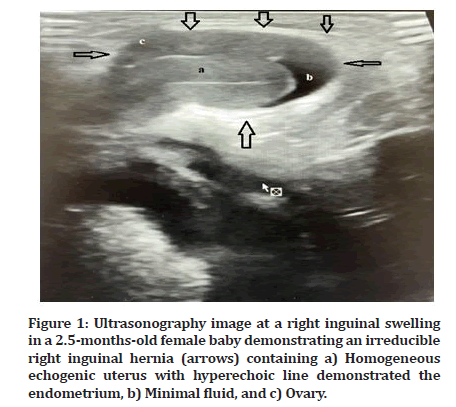
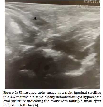
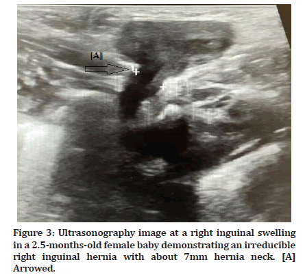
A 2.5-month-old female baby with a symptomatic right inguinal swelling was referred to the radiology department. There was no history of any abnormal conditions related to the vaginal delivery, and there was no history of irritability, redness, pain, vomiting, or signs of an inflammatory process. Ultrasound was used to exclude any abnormal contents, such as the uterus and ovaries. The scanning procedure was performed using high-resolution ultrasound with a linear probe to examine the area of swelling, and the scanning planes differed according to the required area in the swelling. The ultrasound results showed a right inguinal hernia containing the uterus (Figure 1) and ovaries (Figures 1 and 2), which were seen to cross the neck of the hernia. The hernia neck measured approximately 7 mm (Figure 3 Structure A). The hernia was not reducible and contained minimal fluid. The uterus was observed to be not clearly separated from the hernial wall (Figure 1). The vascularity of the reproductive organs was evaluated and showed normal organ perfusion. The patient was referred to the paediatric surgery unit to evaluate the lesion and to assess the patient’s condition, including past and present medical histories. Upon examination, there was an irreducible mass containing the reproductive organs and fluid, as shown on the ultrasound. The patient underwent careful dissection with careful consideration of the possibility of adhesions, and the organs were cleared from the hernial sac. Careful clearance was applied due to the thin hernial wall. Organ reduction and vascular ligation were performed, as well as additional repair of the internal inguinal ring to prevent recurrence. The surgery was successful, and the postoperative course was uneventful. Clinical and radiological follow-up was recommended for one year after the operation.

Figure 1: Ultrasonography image at a right inguinal swelling in a 2.5-months-old female baby demonstrating an irreducible right inguinal hernia (arrows) containing a) Homogeneous echogenic uterus with hyperechoic line demonstrated the endometrium, b) Minimal fluid, and c) Ovary.

Figure 2: Ultrasonography image at a right inguinal swelling in a 2.5-months-old female baby demonstrating a hypoechoic oval structure indicating the ovary with multiple small cysts indicating follicles (A).

Figure 3: Ultrasonography image at a right inguinal swelling in a 2.5-months-old female baby demonstrating an irreducible right inguinal hernia with about 7mm hernia neck. [A] Arrowed.
Discussion
An inguinal hernia is a very common congenital abnormality in infants younger than one year. A few case reports discussing this abnormality have been reviewed in the literature. Hernias containing the uterus are very rare [7,8]. Unusual hernial contents, such as the vermiform appendix, acute appendicitis (Amyand’s hernia) or epiploic appendagitis related to a groin hernia with a massive intrascrotal lipoma (extratesticular), first misdiagnosed as a scrotal hernia, have been reported in cases by Ballas et al. [9].
At six months of foetal development, the processus vaginalis develops as an invagination of the parietal peritoneum. According to gender, it is accompanied by a round ligament or testis and runs through the inguinal canal to the labia majora or scrotum. If patency persists, it is termed the canal of Nuck [10]. A hernia results from an embryological anomaly when there is a failure of partial or complete obliteration of the processus vaginalis in a female [11].
Timely diagnosis is important to exclude solid or cystic components of a hernia and to assess for strangulation and incarceration. Ultrasound is the modality of choice for the characterization and evaluation of inguinal hernias in paediatric patients. However, magnetic resonance imaging is used for diagnosis in adult patients [12]. The ovaries are solid oval structures with a characteristic homogeneous echogenicity and multiple small cysts indicating follicles [11].
Ultrasound is superior to differentiate inguinal hernias from other conditions, including hydrocele, lymphadenopathy, Bartholin gland cyst, infection/ abscess, inguinal gonad, and endometriosis, as well as benign and malignant neoplasms [10].
Surgical intervention is the best management option for hernia repair and treatment in children [10], compared to adult surgery, and it is used for the treatment of symptomatic patients to avoid complications, such as intestinal obstruction and herniated organ ischemia.
Conclusion
Congenital hernia is a common condition seen in babies younger than one year old. This report presents an extremely rare case of a hernia containing the uterus and ovaries that was diagnosed by ultrasound, which is the modality of choice for the diagnosis and differentiation of hernial contents.
Conflict of Interest
They was no conflict of interest.
References
- Karadeniz Cerit K, Ergelen R, et al. Inguinal hernia containing uterus, fallopian tube, and ovary in a premature newborn. Case Rep Pediatr 2015; 16.
- Comella BP, Fortes PO, Salvador RL. Inguinal hernia containing uterus in a newborn: What to do? Pediatr Neonatol 2019; 60:594.
- George EK, Oudesluys-Murphy AM, Madern GC, et al. Inguinal hernias containing the uterus, fallopian tube, and ovary in premature female infants. J Pediatr 2000; 136:696-698.
- Huang CS, Luo CC, Chao HC, et al. The presentation of asymptomatic palpable movable mass in female inguinal hernia. Eur J Pediatr 2003; 162:493-495.
- Cascini V, Lisi G, Di Renzo D, et al. Irreducible indirect inguinal hernia containing uterus and bilateral adnexa in a premature female infant: Report of an exceptional case and review of the literature. J Pediatr Surg 2013; 48:e17-19.
- Clarke S. Pediatric inguinal hernia and hydrocele: An evidence-based review in the era of minimal access surgery. J Laparoendosc Adv Surg Tech A 2010; 20:305-309.
- Thomas AK, Teague CT, Jancelewicz T. Canal of nuck hernia containing pelvic structures presenting as a labial mass. Radiol Case Rep 2018; 13:534-536.
- Jedrzejewski G, Stankiewicz A, Wieczorek AP. Uterus and ovary hernia of the canal of nuck. Pediatr Radiol 2008; 38:1257.
- Ballas K, Kontoulis TH, Skouras CH, et al. Unusual findings in inguinal hernia surgery: Report of 6 rare cases. Hippokratia 2009; 13:169.
- Hamidi H, Rahimi M. Infantile presentation of the canal of Nuck hernia containing uterus and ovary: A case report. Radiol Case Rep 2020; 15:2557-2559.
- Nasser H, King M, Rosenberg HK, et al. Anatomy and pathology of the canal of nuck. Clin Imaging 2018; 51:83-92.
- Robinson A, Light D, Kasim A, et al. A systematic review and meta-analysis of the role of radiology in the diagnosis of occult inguinal hernia. Surg Endosc 2013; 27:11-18.
Indexed at, Google Scholar, Cross Ref
Indexed at, Google Scholar, Cross Ref
Indexed at, Google Scholar, Cross Ref
Indexed at, Google Scholar, Cross Ref
Indexed at, Google Scholar, Cross Ref
Indexed at, Google Scholar, Cross Ref
Indexed at, Google Scholar, Cross Ref
Indexed at, Google Scholar, Cross Ref
Indexed at, Google Scholar, Cross Ref
Indexed at, Google Scholar, Cross Ref
Author Info
Department of Diagnostic Radiology Technology, College of Applied Medical Sciences, Taibah University, Almadinah Almunawwarah, KSAReceived: 21-Oct-2022, Manuscript No. jrmds-22-78100; , Pre QC No. jrmds-22-78100(PQ); Editor assigned: 26-Oct-2022, Pre QC No. jrmds-22-78100(PQ); Reviewed: 09-Nov-2022, QC No. jrmds-22-78100(Q); Revised: 11-Nov-2022, Manuscript No. jrmds-22-78100(R); Published: 18-Nov-2022
