Research - (2022) Volume 10, Issue 3
Knowledge and Awareness of Autogenous Teeth Bone Grafting Material (AutoBt) Among Dental Students-A Survey
Manna A, Gupta A, Tarafder M and Chakraborty S*
*Correspondence: Chakraborty S, Department of Community Medicine, Bankura Sammilani Medical College, India, India,
Abstract
Autogenous tooth bone material is a grafting material prepared from patients extracted teeth. The contents of this grafting material are similar to the contents of the alveolar bone due to some neural crest origin. It plays a positive role in bone remodeling and is used in various procedures. The aim of the study is to assess the knowledge and awareness of autogenous teeth as grafting material among dental students. An online survey was conducted among 100 students consisting of questions on autogenous tooth bone grafting material, its usage, forms, and process to test their knowledge. The result was obtained and respective responses were statistically analyzed in SPSS using descriptive analysis. 57% of the students had knowledge of autogenous tooth bone graft material. 49% answered correctly that it's similar to the contents of alveolar bone. 59% were unaware of its bone remodeling capacity. 68% of the students knew that it can be used effectively for various procedures. 45% were aware and 55% weren't aware of the process of preparation of the grafting material. We can conclude saying that there is insufficient evidence regarding AutoBT as a grafting material even though the majority of students were knowledgeable about it. Therefore, further research is required for definitive conclusions to arrive and make it an easily available and clinically efficient grafting material.
Keywords
Autogenous tooth bone, Grafting material, Bone remodeling, Alveolar bone, Neural crest cells
Introduction
Dental extractions are one of the most common procedures performed when it is no longer possible to restore the function, health and aesthetics of that tooth [1,2]. Post extraction, the alveolar bone undergoes structural and dimensional changes leading to resorption especially on the buccal side of the ridge [3]. Thus, it is important to restore or perform procedures for alveolar ridge preservation. Preservation of alveolar ridge requires ridge augmentation procedures with the help of grafting materials. Autograft, allografts, xenografts and alloplasts are the grafting materials used. Out of all the four, autograft are considered to be the gold standard grafting material because of its osteoconductive, osteoinductive and osteogenic potential [4,5]. It is considered because it has the capacity to maintain stability of the grafted area, ensure rapid revascularization and osteogenesis, rapid healing and no chance of immune rejection due to genetic homogeneity. The other types of grafting materials have the chance of infection, immune rejection, high cost and lack of osteogenic potential [6]. Allografts in particular carry the risk of disease transmission and xenografts and alloplasts only show osteoconduction. Autograft also hold some limitations of limited source, high resorption rates and donor site morbidity [7]. Therefore, alternative grafting materials are required to surpass all these limitations.
Autogenous tooth bone graft material (AutoBT) is a grafting material obtained from patients' own extracted teeth. In 1967, Yeoman and Urist were the first ones to use extracted teeth as a grafting material [8]. This is because the contents of teeth are similar to the content of alveolar bone. Bone and teeth having the same origin from neural crest cells, have similar biochemical composition and histological structure [9]. Alveolar bone contains 65% inorganic and 35% organic material very closely related to dentine with inorganic content of 65-70% which is known to have osteoconductive properties and organic content of 30-35% constituting type 1 collagen [10]. It also consists of growth factors, bone morphogenic protein, non-collagenous proteins like osteocalcin, osteonectin and salioprotein all of which contribute to its osteoinductive property. It plays a positive role in bone remodeling because of the interactions between hydroxyapatite and 3 calcium phosphates - tricalcium phosphate, octacalcium phosphate and amorphous calcium phosphate. The dentin tooth is classified into three different types, each one known to report its potential at different levels. They are the undemineralised dentin(UDD), partially demineralized dentin matrix(PDDM) and demineralized dentin matrix(DDM) [11,12]. The auto tooth graft materials are used in two forms which are particulate and block graft. Block graft shows osteoinductive potential through blood wettability and osteoconductive potential through space maintaining abilities. Similarly, the particles are also based on different sizes of particles, porosities and wettability [13].
extracted teeth are completely cleaned and old restorations, endodontic filling, caries, fibrous soft tissue is removed. Following which, it is sterilized, dried and placed in a chamber to grind into particles. The particles are then immersed in a cleanser solution which is bactericidal in action for 10 minutes. It is demineralized with the help of 2% Nitric acid for 20 minutes following which the dentin is washed with phosphate buffered saline to restore a pH of 7.4 [14]. In this manner, the graft material is ready to be used for ridge augmentation procedures, ridge splitting, sinus lifting, guided bone regeneration etc. This method makes use of extracted teeth and is known to give positive results. This can surpass the limitations of other grafting materials and more awareness can be created for people to use this. The aim of this study is to assess the knowledge and awareness of Autogenous tooth bone graft material among dental students.
Materials and Methodology
The study was conducted among a total of 100 dental students of the undergraduate and postgraduate programme through an online survey. A self-administered questionnaire was given to these students to assess their knowledge and awareness on autogenous teeth as a grafting material, processing, forms in which it can be used, advantages, disadvantages and complications. It also was designed to analyze their understanding and knowledge on grafting materials and requirements of an ideal grafting material. The data was collected and results were obtained through descriptive analysis in the form of bar charts with the help of SPSS version 23.0.
Results and Discussion
The survey was conducted among 100 students where 55 were females and 45 were males. The results of the individual questions are shown below in the form of bar charts. Figure 1 shows that 57% of the students had knowledge of autogenous tooth bone graft material.
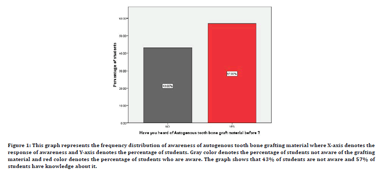
Figure 1: This graph represents the frequency distribution of awareness of autogenous tooth bone grafting material where X-axis denotes the response of awareness and Y-axis denotes the percentage of students. Gray color denotes the percentage of students not aware of the grafting material and red color denotes the percentage of students who are aware. The graph shows that 43% of students are not aware and 57% of students have knowledge about it.
When questioned about the contents of teeth similar to which kind of tissue, 49% answered correctly that it's similar to the contents of alveolar bone (Figure 2). Figure 3 showed that 53% of the students felt that posterior teeth are more suitable to be used as grafting material than anterior teeth. 67% of the students chose the particulate type of graft, 22% chose block grafting and 11% chose to use a mixture of block and particulate grafting as seen in Figure 4.
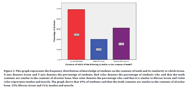
Figure 2:This graph represents the frequency distribution of knowledge of students on the contents of teeth and its similarity to which tissue. X-axis denotes tissue and Y-axis denotes the percentage of students. Red color denotes the percentage of students who said that the teeth contents are similar to the contents of alveolar bone, blue color denotes the percentage who said that it is similar to fibrous tissue and violet color represents tendon and muscle. The graph shows that 49% of students said that the teeth contents are similar to the contents of alveolar bone, 20% fibrous tissue and 31% tendon and muscle.
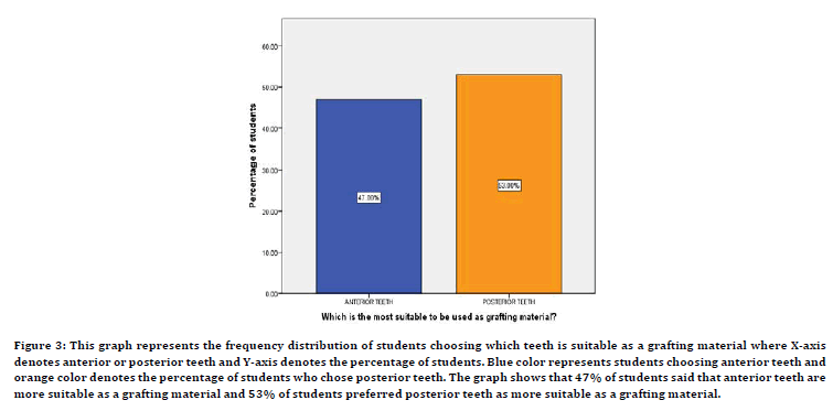
Figure 3:This graph represents the frequency distribution of students choosing which teeth is suitable as a grafting material where X-axis denotes anterior or posterior teeth and Y-axis denotes the percentage of students. Blue color represents students choosing anterior teeth and orange color denotes the percentage of students who chose posterior teeth. The graph shows that 47% of students said that anterior teeth are more suitable as a grafting material and 53% of students preferred posterior teeth as more suitable as a grafting material.
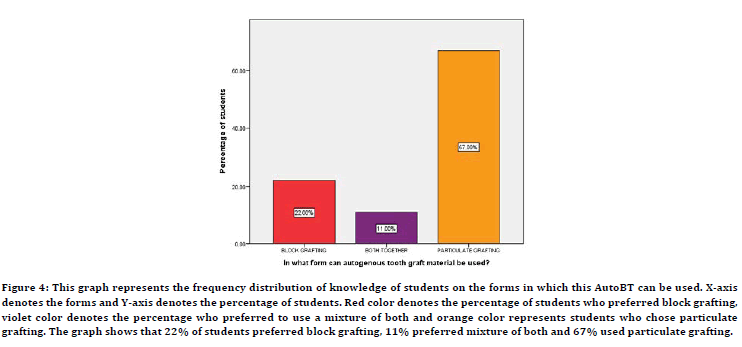
Figure 4:TThis graph represents the frequency distribution of knowledge of students on the forms in which this AutoBT can be used. X-axis denotes the forms and Y-axis denotes the percentage of students. Red color denotes the percentage of students who preferred block grafting, violet color denotes the percentage who preferred to use a mixture of both and orange color represents students who chose particulate grafting. The graph shows that 22% of students preferred block grafting, 11% preferred mixture of both and 67% used particulate grafting.
Figure 5 depicted the frequency of students who were aware of the osteoinductive potential and we see that 59% were unaware of its bone remodeling capacity. 68% of the students knew that AutoBT can be used for ridge augmentation, ridge splitting and socket preservation as shown in Figure 6. 22% of the students, as depicted in Figure 7, chose to not mix autogenous teeth bone graft with other grafting materials. Students were also asked if they had knowledge about the process of deriving grafting material from extracted teeth and the response as depicted in Figure 8 is that 45% were aware and 55% weren't aware of the process.
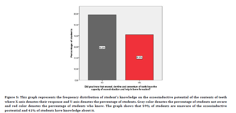
Figure 5:This graph represents the frequency distribution of student's knowledge on the osseoinductive potential of the contents of teeth where X-axis denotes their response and Y-axis denotes the percentage of students. Gray color denotes the percentage of students not aware and red color denotes the percentage of students who know. The graph shows that 59% of students are unaware of the osseoinductive postential and 41% of students have knowledge about it.
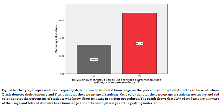
Figure 6:This graph represents the frequency distribution of students' knowledge on the procedures for which AutoBT can be used where X-axis denotes their response and Y-axis denotes the percentage of students. Gray color denotes the percentage of students not aware and red color denotes the percentage of students who knew about its usage in various procedures. The graph shows that 32% of students are unaware of the usage and 68% of students have knowledge about the multiple usages of the grafting material.
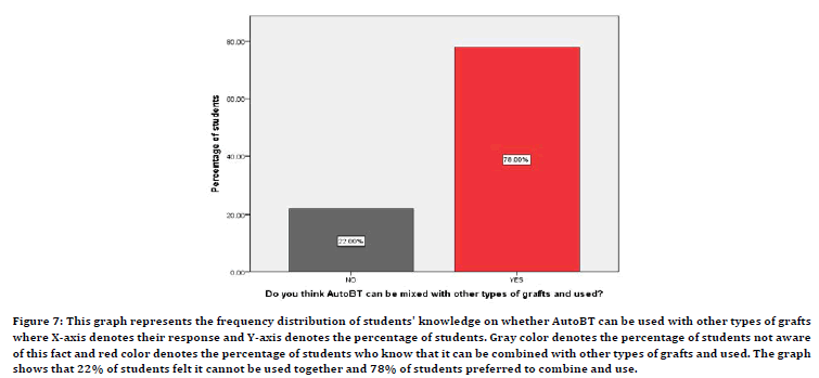
Figure 7:This graph represents the frequency distribution of students' knowledge on whether AutoBT can be used with other types of grafts where X-axis denotes their response and Y-axis denotes the percentage of students. Gray color denotes the percentage of students not aware of this fact and red color denotes the percentage of students who know that it can be combined with other types of grafts and used. The graph shows that 22% of students felt it cannot be used together and 78% of students preferred to combine and use.
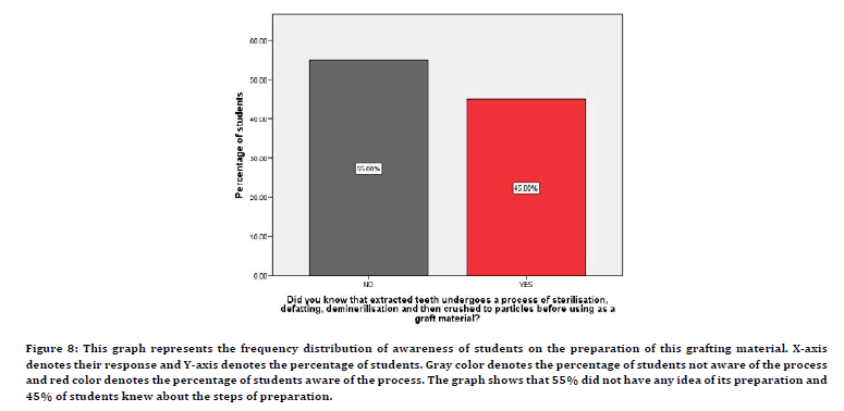
Figure 8:This graph represents the frequency distribution of awareness of students on the preparation of this grafting material. X-axis denotes their response and Y-axis denotes the percentage of students. Gray color denotes the percentage of students not aware of the process and red color denotes the percentage of students aware of the process. The graph shows that 55% did not have any idea of its preparation and 45% of students knew about the steps of preparation.
Discussion
Our team has contributed high quality publications through their extensive knowledge and remarkable research experience [15–34]. Auto BT has an elaborate collagen structure proving an ideal environment for differentiation and proliferation to initiate bone formation. Murata et al reported that demineralised teeth generated bone on research due to similarity with bone components [35]. This was in accordance with our study where 49% of the students reported that the contents of teeth are similar to that of bone as shown in Figure 2. It is known to be biocompatible with no chance of infection and rejection [36]. Figure 1 shows 57% of the students were aware of this and considered autogenous tooth bone as a grafting material. The extracted teeth undergo a burnout method to remove organic material and form particulate dentin [37]. The high temperature majorly affects the hydroxyapatite crystals and b-TCP according to Kim et al. The root area consists of dentin and cementum showing low crystallinity whereas the crown part contains more enamel components showing high crystallinity. Moreover, posterior teeth are more preferred as grafting material than anterior teeth because of increased contents as compared to anterior teeth thereby showing similarity to our study where 53% chose posteriors as shown in Figure 3 [38,39]. Crown and root show different healing mechanisms due to differences in their crystallinity. Low crystalline carbonic apatites show good osteoconductive effect whereas small crystalline sizes of HA help in good biodegradation because of high solubility [40,41]. Figure 5 depicts that 59% of students were unaware of enamel, dentin and cemnutm having osseoinduction potential and for bone formation. Ike and Urist in his study proved that enamel containing BMP-2 has osteoinductive capacity leading to bone formation [42].
63% of students (Figure 6) had knowledge about the usage of the AutoBT in various procedures and wished to use them more often rather than using other synthetic grafting materials. In various clinical studies it has been found that AutoBT is used in guided bone regeneration, ridge augmentation, ride splitting and socket preservation [43]. It has also been used to treat grade II and III furcation defects where it demonstrated a reduction in vertical and horizontal bone depth [44]. Another study showed the usage of this grafting material in sinus floor elevation procedures compared to Bio-Oss which did not show any significant difference between the two [45]. Figure 4 showed that 67% of students preferred particulate grafting, 22% chose block grafting and the remaining 11% chose to use both together to graft. Some studies have shown that there is no significant difference in the volumetric reduction between the two [46]. Moreover, there was no crestal bone loss also seen. Contrary to this study, crestal bone loss was reported in patients where particulate grafting was used rather than blocks. There have been protocols suggesting the usage of AutoBt with other types of grafts. Lee et al, in his study combined it with xenobone, allobone and synthetic bone to decrease the resorption and increase implant stability confirming that AutoBT along with other types of grafts helps improve the implant stability gradually [47]. 78% of the students in our study (Figure 7) also supported this fact and are in compliance with the study that a mixture of grafting materials gives better results.
Although AutoBT has been proven to give excellent results due to its several advantages, it holds few limitations as well. The preparation of this grafting material is time consuming and isn't possible chairside. Ideal particle size for new bone formation is 300-1200 um and the size differs for maxilla and mandible [14,48]. Along with this, its limited availability, healing mechanisms and bone resorption also is to be worked upon.
Conclusion
Within the limitations of the study, we conclude that the majority of the students were aware and had knowledge about autogenous tooth bone grafting. Nonetheless, it still requires advancement with the help of future technology and ways of improving its clinical efficacy to make it a commercially available grafting material. Since there is a scarcity of studies, evidence and standardized protocol, students are unsure of using the material more often. Therefore, more research, knowledge and awareness is required for a definitive conclusion to gain more efficacy for this grafting material.
Conflict of Interest
The authors declare that there are no conflicts of interest in the present study.
Acknowledgement
We would like to thank our college and management for their constant support in completing this research work.
References
- Avila-Ortiz G, Elangovan S, Kramer KWO, et al. Effect of alveolar ridge preservation after tooth extraction. J Dent Res 2014; 93:950–958.
- Van der Weijden F, Dell'Acqua F, Slot DE. Alveolar bone dimensional changes of post‐extraction sockets in humans: A systematic review. J Clin Periodont 2009; 36:1048-58.
- Tan WL, Wong TL, Wong MC, et al. A systematic review of post‐extractional alveolar hard and soft tissue dimensional changes in humans. Clin Oral Implants Res 2012; 23:1-21.
- Jambhekar S, Kernen F, Bidra AS. Clinical and histologic outcomes of socket grafting after flapless tooth extraction: A systematic review of randomized controlled clinical trials. J Prosthet Dent 2015; 113:371–82.
- Ramanauskaite A, Sahin D, Sader R, et al. Efficacy of autogenous teeth for the reconstruction of alveolar ridge deficiencies: A systematic review. Clin Oral Investig 2019; 23:4263–87.
- Quattlebaunr JB, Mellonig JT, Hensel NF. Antigenicity of freeze-dried cortical bone allograft in human periodontal osseous defects. J Periodontol 1988; 59:394–397.
- Sogal A, Tofe AJ. Risk assessment of bovine spongiform encephalopathy transmission through bone graft material derived from bovine bone used for dental applications. J Periodontol 1999; 70:1053–63.
- Pentapati KC, Smriti K, Bhattacharjya C, et al. Tooth derived bone graft material. World J Dent 2016; 7:32–35.
- https://www.elsevier.com/books/orbans-oral-histology-and-embryology/kumar/978-81-312-4033-5
- Gual-Vaqués P, Polis-Yanes C, Estrugo-Devesa A, et al. Autogenous teeth used for bone grafting: A systematic review. Med Oral Patol Oral Cirugia Bucal 2018; 23:e112.
- Kim YK, Kim SG, Bae JH, et al. Guided bone regeneration using autogenous tooth bone graft in implant therapy. Implant Dent 2014; 23:138–43.
- Cenicante J, Botelho J, Machado V, et al. The use of autogenous teeth for alveolar ridge preservation: A literature review. App Sci 2021; 11: 1853.
- Wadhwa P, Lee JH, Zhao BC, et al. Microcomputed Tomography and histological study of bone regeneration using tooth biomaterial with BMP-2 in rabbit calvarial defects. Scanning 2021; 2021:6690221.
- Minamizato T, Koga T, Takashi I, et al. Clinical application of autogenous partially demineralized dentin matrix prepared immediately after extraction for alveolar bone regeneration in implant dentistry: A pilot study. Int J Oral Maxillofac Surg 2018; 47:125–32.
- Ramesh A, Varghese S, Jayakumar ND, et al. Comparative estimation of sulfiredoxin levels between chronic periodontitis and healthy patients: A case-control study. J Periodontol 2018; 89:1241–1248.
- Paramasivam A, Priyadharsini JV, Raghunandhakumar S, et al. A novel COVID-19 and its effects on cardiovascular disease. Hyper Res 2020; 43:729–30.
- Gokila S, Gomathi T, Vijayalakshmi K, et al. Development of 3D scaffolds using nanochitosan/silk-fibroin/hyaluronic acid biomaterials for tissue engineering applications. Int J Biol Macromol 2018; 120:876-85.
- Del Fabbro M, Karanxha L, Panda S, et al. Autologous platelet concentrates for treating periodontal infrabony defects. Cochrane Database Syst Rev 2018; 11:CD011423.
- Paramasivam A, Vijayashree Priyadharsini J. MitomiRs: New emerging microRNAs in mitochondrial dysfunction and cardiovascular disease. Hyper Res 2020; 43:851-853.
- Jayaseelan VP, Arumugam P. Dissecting the theranostic potential of exosomes in autoimmune disorders. Cell Mol Immunol 2019; 16:935.
- Vellappally S, Al Kheraif AA, Divakar DD, et al. Tooth implant prosthesis using ultra low power and low cost crystalline carbon bio-tooth sensor with hybridized data acquisition algorithm. Computer Commun 2019; 148:176–184.
- Vellappally S, Al Kheraif AA, Anil S, et al. Analyzing relationship between patient and doctor in public dental health using particle memetic multivariable logistic regression analysis approach (MLRA2). J Med Systems 2018; 42.
- Varghese SS, Ramesh A, Veeraiyan DN. Blended module‐based teaching in biostatistics and research methodology: A retrospective study with postgraduate dental students. J Dent Educ 2019; 83:445-50.
- Venkatesan J, Singh SK, Anil S, et al. Preparation, characterization and biological applications of biosynthesized silver nanoparticles with chitosan-fucoidan coating. Molecules 2018; 23:1429.
- Alsubait SA, Al Ajlan R, Mitwalli H, et al. Cytotoxicity of different concentrations of three root canal sealers on human mesenchymal stem cells. Biomol 2018; 8:68.
- Venkatesan J, Rekha PD, Anil S, Bhatnagar I, et al. Hydroxyapatite from cuttlefish bone: Isolation, characterizations, and applications. Biotechnol Bioprocess Eng 2018; 23:383-93.
- Vellappally S, Al Kheraif AA, Anil S, et al. IoT medical tooth mounted sensor for monitoring teeth and food level using bacterial optimization along with adaptive deep learning neural network. Measurement 2019; 135:672-7.
- PradeepKumar AR, Shemesh H, Nivedhitha MS, et al. Diagnosis of vertical root fractures by cone-beam computed tomography in root-filled teeth with confirmation by direct visualization: A systematic review and meta-analysis. J Endod 2021; 47:1198-214.
- Ramani P, Tilakaratne WM, Sukumaran G, et al. Critical appraisal of different triggering pathways for the pathobiology of pemphigus vulgaris-A review. Oral Dis 2021.
- Ezhilarasan D, Lakshmi T, Subha M, et al. The ambiguous role of sirtuins in head and neck squamous cell carcinoma. Oral Dis 2021.
- Sarode SC, Gondivkar S, Sarode GS, et al. Hybrid oral potentially malignant disorder: A neglected fact in oral submucous fibrosis. Oral Oncol 2021; 121:105390.
- Kavarthapu A, Gurumoorthy K. Linking chronic periodontitis and oral cancer: A review. Oral Oncol 2021; 121:105375.
- Vellappally S, Abdullah Al-Kheraif A, Anil S, et al. Maintaining patient oral health by using a xeno-genetic spiking neural network. J Ambient Intell Humaniz Comput 2018; 1-9.
- Aldhuwayhi S, Mallineni SK, Sakhamuri S, et al. Covid-19 knowledge and perceptions among dental specialists: a cross-sectional online questionnaire survey. Risk Manage Healthcare Policy 2021; 14:2851.
- Murata M, Akazawa T, Takahata M, et al. Bone induction of human tooth and bone crushed by newly developed automatic mill. J Ceramic Society Japan 2010; 118:434-7.
- Ellegaard B, Karring T, Davies R, Löe H. New attachment after treatment of intrabony defects in monkeys. J Periodontol 1974; 45:368–77.
- Kim MG, Lee JH, Kim GC, et al. The effect of autogenous tooth bone graft material without organic matter and type I collagen treatment on bone regeneration. Maxillofac Plastic Recon Surg 2021; 43:1-3.
- Kim YK, Kim SG, Byeon JH, et al. Development of a novel bone grafting material using autogenous teeth. Oral Surg Oral Med Oral Pathol Oral Radiol Endodontol 2010; 109:496-503.
- Kim YK, Jun SH, Um IW, et al. Evaluation of the healing process of autogenous tooth bone graft material nine months after sinus bone graft: Micromorphometric and histological evaluation. Maxillofac Plastic Reconst Surg 2013; 35:310-315.
- Lu J, Descamps M, Dejou J, et al. The biodegradation mechanism of calcium phosphate biomaterials in bone. J Biomed Mater Res 2002; 63:408–12.
- Fulmer MT, Ison IC, Hankermayer CR, et al. Measurements of the solubilities and dissolution rates of several hydroxyapatites. Biomater 2002; 23:751-755.
- Ike M, Urist MR. Recycled dentin root matrix for a carrier of recombinant human bone morphogenetic protein. J Oral Implantol 1998; 24:124-32.
- Reddy GV, Abhinav A, Malgikar S, et al. Clinical and radiographic evaluation of autogenous dentin graft and demineralized freeze-dried bone allograft with chorion membrane in the treatment of Grade II and III furcation defects-: A randomized controlled trial. Indian J Dent Sci 2019; 11:83.
- Jun SH, Ahn JS, Lee JI, et al. A prospective study on the effectiveness of newly developed autogenous tooth bone graft material for sinus bone graft procedure. J Adv Prosthod 2014; 6:528-38.
- Dasmah A, Thor A, Ekestubbe A, et al. Particulate vs. block bone grafts: Three-dimensional changes in graft volume after reconstruction of the atrophic maxilla: A 2-year radiographic follow-up. J Craniomaxillofac Surg 2012; 40:654–659.
- Lee JY, Kim YK, Yi YJ, et al. Clinical evaluation of ridge augmentation using autogenous tooth bone graft material: case series study. J Korean Assoc Oral Maxillofac Surg 2013; 39:156.
- Binderman I, Hallel G, Nardy C, et al. A novel procedure to process extracted teeth for immediate grafting of autogenous dentin. J Interdiscipl Med Dent Sci 2014; 2:2.
Indexed at, Google Scholar, Cross Ref
Indexed at, Google Scholar, Cross Ref
Indexed at, Google Scholar, Cross Ref
Indexed at, Google Scholar, Cross Ref
Indexed at, Google Scholar, Cross Ref
Indexed at, Google Scholar, Cross Ref
Indexed at, Google Scholar, Cross Ref
Indexed at, Google Scholar, Cross Ref
Indexed at, Google Scholar, Cross Ref
Indexed at, Google Scholar, Cross Ref
Indexed at, Google Scholar, Cross Ref
Indexed at, Google Scholar, Cross Ref
Indexed at, Google Scholar, Cross Ref
Indexed at, Google Scholar, Cross Ref
Indexed at, Google Scholar, Cross Ref
Indexed at, Google Scholar, Cross Ref
Indexed at, Google Scholar, Cross Ref
Indexed at, Google Scholar, Cross Ref
Indexed at, Google Scholar, Cross Ref
Indexed at, Google Scholar, Cross Ref
Indexed at, Google Scholar, Cross Ref
Indexed at, Google Scholar, Cross Ref
Indexed at, Google Scholar, Cross Ref
Indexed at, Google Scholar, Cross Ref
Indexed at, Google Scholar, Cross Ref
Indexed at, Google Scholar, Cross Ref
Indexed at, Google Scholar, Cross Ref
Indexed at, Google Scholar, Cross Ref
Indexed at, Google Scholar, Cross Ref
Indexed at, Google Scholar, Cross Ref
Indexed at, Google Scholar, Cross Ref
Indexed at, Google Scholar, Cross Ref
Indexed at, Google Scholar, Cross Ref
Indexed at, Google Scholar, Cross Ref
Indexed at, Google Scholar, Cross Ref
Indexed at, Google Scholar, Cross Ref
Indexed at, Google Scholar, Cross Ref
Indexed at, Google Scholar, Cross Ref
Indexed at, Google Scholar, Cross Ref
Indexed at, Google Scholar, Cross Ref
Indexed at, Google Scholar, Cross Ref
Indexed at, Google Scholar, Cross Ref
Indexed at, Google Scholar, Cross Ref
Author Info
Manna A, Gupta A, Tarafder M and Chakraborty S*
1Department of Periodontics, Saveetha Dental College and Hospitals, Saveetha Institute of Medical and Technical Sciences, Saveetha University, Chennai-77, Tamil Nadu, India2India
3Department of Community Medicine, Bankura Sammilani Medical College, India, India
4Department of Community Medicine, Bankura Sammilani Medical College, India, India
Citation: Manna A, Gupta A, Tarafder M, Chakraborty S, Knowledge and Awareness of Autogenous Teeth Bone Grafting Material (AutoBt) Among Dental Students-A Survey, J Res Med Dent Sci, 2022, 10 (3):150-155.
Received: 13-Feb-2022, Manuscript No. JRMDS-22-54340; , Pre QC No. JRMDS-22-54340 (PQ); Editor assigned: 15-Feb-2022, Pre QC No. JRMDS-22-54340 (PQ); Reviewed: 01-Mar-2022, QC No. JRMDS-22-54340; Revised: 14-Mar-2022, Manuscript No. JRMDS-22-54340 (R); Published: 16-Mar-2022
