Review Article - (2023) Volume 11, Issue 8
Mandibular Molar Protraction
Shambhavi S Moharil*, Kamble Ranjit and Renuka Talla
*Correspondence: Dr. Shambhavi S Moharil, Department of Orthodontics, Datta Meghe Institute of Medical Sciences, Wardha, India, Email:
Abstract
Many orthodontists worry about the spacing in the mandibular arch whether it is due to second molar extraction or due to the missing second premolar. Missing mandibular molar can cause problem such as posterior spacing. If the clinician wants to go for a prosthetic outlook, then there should be left an exact amount of space in the arch and alveolar ridge should be kept unharmed. There are conditions in which the patient desires for a whole set of natural dentitions as well as cases where molar mesialization can correct the malocclusion and simultaneously improve the profile of the patient. There are various appliances which make this mechanism possible which include loop mechanics such as cherryloop, running loop, T loop, V bends and tip back, sliding bends and bars, etc. Recent advances in TADS increased the scope of molar protraction in cases where the retraction of anteriors is not advocated. Without the retraction of the anteriors and premolars, protraction of mandibular second and third molar becomes even more challenging hence TADS can be beneficial. For achieving molar protraction that to mandibular several approaches are being recognized corticotomy assisted molar protraction, PAOO. With this mesialization of the 2nd molar even the impacted 3rd molar can be given space in the arch without surgical intervention. With this treatment natural dentition is achieved i.e., there will be no need of prosthetic implants and bridges for treating cases of missing posteriors. This review articlefocuses on the indications of the molar protraction and the various modalities with which we can achieve mesialization of the posterior segment.
Keywords
Molar protraction, Space closure, Edentulous arch, Alveolar ridge, Molar mesialization
Introduction
Molar distalization has been in the limelight since ages while molar protraction has never been the topic of discussion. Missing mandibular molar can cause problem such as posterior spacing. This problem can be treated only if the diagnosis is early i.e., during the mixed dentition period. Dental anomalies in tooth number, shape, structure and position influences treatment planning by causing malocclusion and other problems [1]. Congenitally missing third molars had the prevalence of approximately 16.3%. Impacted third molars had the prevalence of about 9.7%. The orthodontists usually choose one of the two options as their treatment plan and this decides whether to keep the molar or to extract it. If the spacing is planned for the prosthetic treatment, the orthodontist is required to create that much space as well as keep the alveolar ridge safe and sound. If the orthodontist chooses to get rid of the spacing, he/she must protract the molar [2].
If mandibular first molar is lost, the closure of spacing is best done by reciprocal movement of the all the teeth present. Roberts et al. used endosseous implants to close the space by mesial movement of the mandibular second and third molar of the same quadrant. It is very difficult to protract the mandibular posterior tooth especially molar due to its increased density. Without the retraction of the anteriors and premolars, protraction of mandibular second and third molar becomes even more challenging [3].
Cortical plates both buccal and lingual play a very crucial role in the treatment hence any anomaly or collapse may lead to complications. Intraoral skeletal anchorage like Temporary Anchorage Devices (TADs), miniscrews, plates provide good and rapid results. After second molar protraction, the problem of impacted third molar is solved automatically as there is scope of natural eruption of the third molar in an upright position without any special treatment [4]. Molar protraction if carried out successfully then there is proper closure of the missing posterior spaces and hence implants and bridges will no longer be required. By engaging the third molar the value, significance and success of the treatment is witnessed. Due to aging the periodontal and root resorption problems become more significant and this causes difficulty in molar protraction [5].
Literature Review
Why is molar protraction difficult in mandible?
There is structural difference in both the maxillary arch and mandibular arch hence; there is reduced support in the mandibular arch. In maxilla, the molar region has approximately 1.5 mm of buccal cortical plate thickness, whereas mandible has 2 mm thickness. As the molar protraction increases there is simultaneous decrease of the cortical thickness [6].
Classification of molar protraction
Molar protraction can be classified on the basis of the missing area:
• U-6
• L-6
• L-E
The amount of posterior teeth movement:
• Pure anterior teeth retraction
• Reciprocal traction
• Pure protraction of posterior teeth
Indications
• Condition where there is minimal overjet with
generalised spacing in cases with lass I, II and III
malocclusions.
• Cases where the premolar extraction was carried out
and the extraction spaces are left after retraction of
the anteriors in cases of class I type 1 malocclusion.
• When permanent molars which are grossly carious,
the 2nd molars are to be mesialized.
• Class II molar relationships where mandible is
retrognathic.
• When the molar correction is required in class III
malocclusion.
• Loss of anchorage during orthodontic treatment.
• Excessive lower anterior facial height.
• In open bite cases where the mesialization of
mandibular molar into the extraction site aids in
closure of bite.
• Cases where the second premolar extraction was
carried out in order to correct the molar relation like
end on relation or full cusp class II molar relation due
retrognathism of mandible.
Contraindications
• Cases with reduced lower anterior facial elevation
• Skeletal deep bite
• Horizontal growth pattern
Discussion
Intra oral appliances
Lingual elastic tied to the archwire: Miniscrew can create posterior crossbite and open bite due to direct protraction. To avoid these obstacles, following steps should be given attention (Figure 1).
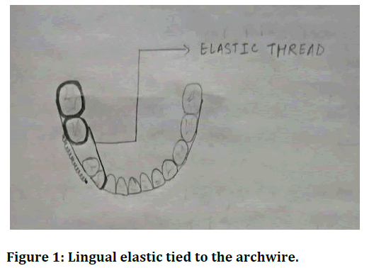
Figure 1: Lingual elastic tied to the archwire.
• Mesialization with an optimum lingual pressure, such
as an elastic thread tie from lingual surface of molar
to the archwire, for preventing rotation of the
anteriors, incisors and canines must be ligated.
• Arch expansion can be minimized by involving the
second molar into the archwire.
• Molar can rotate buccally, so to prevent that
rectangular archwire is used.
• With the help of auxiliary slots, to retract the tooth at
its centre of resistance a hook placed bucally can be
produced from fragment of wire.
Sliding band on lingual molar: Whenever we protract an extreme tooth into the arch, there is a fear of that tooth going into the crossbite, hence it is of a great importance to have a balanced lingual force for that tooth. The lingual arch has a 0.040 wire soldered on the opposite side of the lone molar on its band (Figure 2) [7,8].
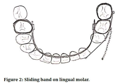
Figure 2: Sliding band on lingual molar.
Sliding band and bar: A 0.032 stainless steel wire which is introduced on both the sides of the arch in the tubes is soldered anteriorly to the second premolar band. Soldering of the hooks is carried out near the CR of the molar and premolar so as to apply chain [9]. There is a segment on the buccal aspect of the premolar where a 0.21 × 0.25 rigid wire can be attached to gain an indirect support from a mini-implant between the lower premolars [10]. Cementing of the appliance is done and from the buccal aspect of the mini-implant a rigid stainless steel power arm is bent, which is engaged in the premolar tube and cinched. Splinting of the stainlesssteel fragment is achieved by using the flowable type of composite onto the mini-implant (Figure 3).
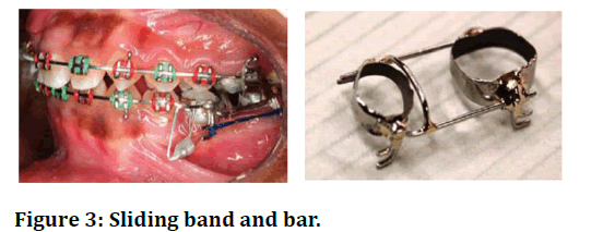
Figure 3: Sliding band and bar.
The push-pull technique: Traditionally, to avoid hindrance of the molar protraction, a miniscrew is advised to be placed mesial to the edentulous space. As a substitute, a very good option is usage of TADs. This technique works by placing the TADs within the edentulous area to support the second molar from behind, with the help of an open-coil spring the tooth is moved in front of it, complete space closure is gained as the spring moves the tooth (Figure 4) [11].
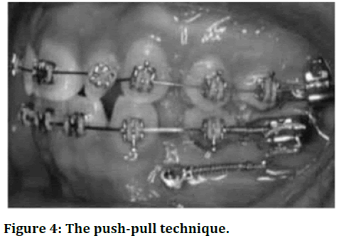
Figure 4: The push-pull technique.
Following are the reasons why we prefer the push-pull technique over other protraction methods:
• Simplifies miniscrew insertion
• Ensures adequate bone stock
• Minimal root perforation
• Auxiliary is prevented from crossing the canine
eminence
• For efficient multitooth protraction two active forces
are applied (open-coil spring and nickel-titanium coil
spring)
Class 2 elastics: When class II elastics are given, due to its properties such as mesialization of mandibular molars, closure of space is possible. The elastics also prevent the mesial tipping of the mandibular molars and help in bodily movement. Class II elastics are quite effective when it comes to mandibular molar protraction. The retruded mandible is quite common in strong facial patterns and here the class II elastics can help achieve overall facial balance (Figure 5) [12].
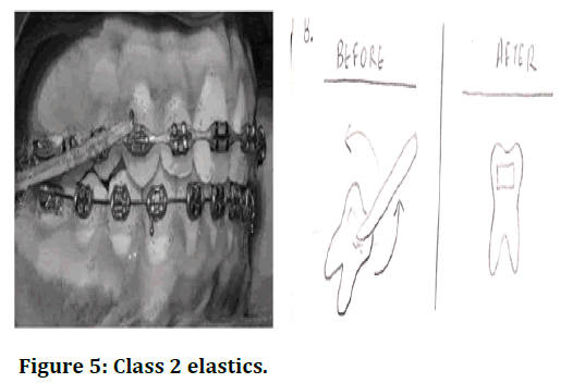
Figure 5: Class 2 elastics.
Sved inclined plane: The sved inclined plane is positioned in the maxillary arch. The role of this inclined plane is such that it brings the mandibular towards in the desired position. The lower anteriors are positioned ahead of the inclined plane which creates a force on upper anteriors in forward and upward direction leading to proclination. This proclination can be prevented by incisal capping. The forward placement of the mandible creates a restraining force on maxillary arch which aids in anchorage reinforcement in the upper arch. In the mandibular arch as the bite plane disoccludes the dentition. The mesial movement of the posterior teeth can be carried out faster as there are no occlusal interferences (Figure 6) [13].
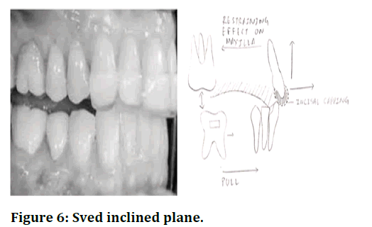
Figure 6: Sved inclined plane.
V-bends tip-backbends: Elicited movement of the teeth without friction or erosion or attrition. The force system generated by V bend in a straight wire was studies by means of the principles for small deflection of a beam and a model developed for the forces and moments. The positioning between “geometries” and V bends which is developed between straight wire and angulated brackets is drawn. It is very difficult to protract the lower molar because of presence of various vital structures such as bulky buccolingual root of mandibular molar, cortical bone and thickness of the bone mesial to the first molar is decreased because to deficit buccolingual girth of the second premolar. By using “V” bend mechanic the patient feels lesser pain and this is very advantageous (Figure 7) [14].
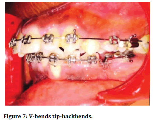
Figure 7: V-bends tip-backbends.
Cherry loop: It is made of strong and stable 0.17 × 0.25 stainless steel wire. This wire is sufficiently flexible to enter without any distortion in a 0.22 × 0.28 molar tube. Rouland plier is used to give it a bend. The tube must be placed at half the distance away so as to separate the bracket of the mandibular first premolar from the molar tube of the first molar. Once the protraction of the molar begins, the loop should cover 50% of the distance. This can be achieved by giving a V bent at the distal aspect of the canine which will shorten the wire (Figure 8) [15].
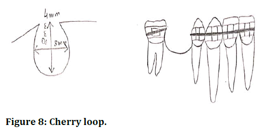
Figure 8: Cherry loop.
Running loop: It is a very simple, easy and efficient technique for terminating the gap or space without causing any tooth movements such as tipping and rotation mesially or lingually. This is achieved by promoting uprighting and mesialization of molars which go hand in hand commonly known as walking of molars. It is made up of stainless-steel wire 0.018 × 0.025. The diameter of the helical running loop measures about 3 mm. About 5 mm of the distance is maintainable within the buccal tube and the running loop (Figure 9) [16].
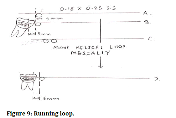
Figure 9: Running loop.
T loop: Hoenigl et al modified the original design (by Brustone) in the year 1995 by fabricating a longer vertical arm and applied force reduction. T loop can be fabricated by using SS wire or TMA wire. The T loop measures total length of 10 mm where the height is approx. 2 mm, the mesial and the distal leg measures 2 mm and 5 mm respectively. Due to simultaneous uprighting and mesialization of molars the T loop gets activated causing the translatory movement of teeth (Figure 10) [17].
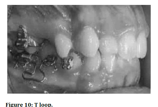
Figure 10: T loop.
Piezocision: The major disadvantage of the orthodontic treatment is its duration of treatment; hence many adults go for a prosthetic approach and prefer to invest money rather than time. But many treatments and methods have come forward to lessen this time interval between the commencement and result of the treatment. There are two types to accelerate the tooth mobility i.e., surgical approach and a nonsurgical approach. Piezocision is one of the latest surgical methods used to accelerate tooth movement Dibart et al were the first to describe this minimally invasive procedure that combined microincisions limited to the buccal side creating small cuts into the bone using a piezotome (Figure 11) [18].
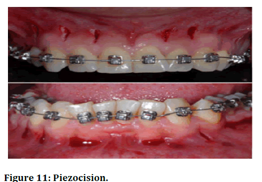
Figure 11: Piezocision.
Surgically assisted molar protraction: Periodontally Accelerated Osteogenic Orthodontics (PAOO) is an advanced method which involves corticotomies and a few selective bone allografts and this further enhances the rate of tooth mesialization by increasing alveolar bones rate of replacement and further reducing the bone thickness. PAOO approach is used for bilateral molar protraction using mini-screws forsupport. Corticotomy was carried out with the help of TADs in a very famous case, but this surgical intervention was performed on the maxillary first molar (Figure 12) [19].
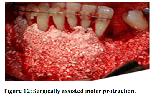
Figure 12: Surgically assisted molar protraction.
Dual geometry archwire (Hills): This has been designed for the sliding mechanics in the posterior segment with the help of a polished round posterior segment. The archwire is built with a strong and stable stainless-steel wire. The hills archwire is available in 2 sizes: 0.018 × 0.018 inches anterior with 0.018-inch round posterior for the 0.018 slot and 0.021 × 0.021 inches anterior with 0.020-inch round posterior for the 0.022 slot.
Bidimensional system: For incisors small brackets of slot number 0.018 × 0.025 were used for a good and perfect fitting three-dimensional control and much larger slot number of 0.022 × 0.028 were used for the posterior teeth. Thus, giving it a loose fit due to exertion of force due to lever mechanics.
Fixed functional appliances: Twin force bite corrector can be to simultaneously correct class II malocclusion and serve as an anchorage for protraction of a mandibular molar into the edentulous space. There are two parallel 15 mm cylindrical appliance which provide shelter to nickel titanium coil springs and there is plunger in each cylinder at opposite ends (Figure 13).
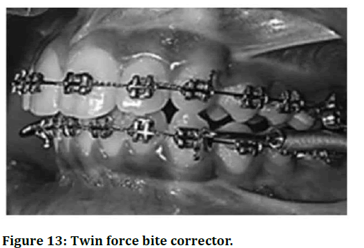
Figure 13: Twin force bite corrector.
Mandibular protraction appliance: It is a fixed functional appliance, which is brand new innovation, it is rigid and noncompliant and it there is anterior involvement of the mandible and there is correction of the class II antero-posteriorly balance. This is applicable and useful in mandibular molar protraction patients. To correct the class II malocclusion four types of protraction appliances are given: MPA I, MPA II, III and IV (Figure 14).
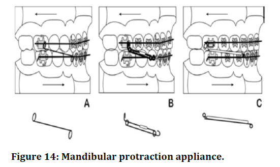
Figure 14: Mandibular protraction appliance.
Braking mechanics
The brakes reverse the anchorage site from the posterior to the anterior segment by permitting only bodily movement of the anterior teeth. This is achieved by using braking mechanics such as springs and torquing component on the anterior teeth. One or more forms of brakes are applied depending on the braking needs. The commonly used brakes are as follows.
Breaking springs: These are passive uprighting springs made in 0.018 wire, which almost fill the bracket channel.
Torquing auxiliaries: A two spur or four spur auxiliary, which is activated to the desired extent or a MAA design in 0.010 or 0.011 size can also be used as a braking mechanism along with a strong base wire (0.020 or 0.018) (Figure 15).
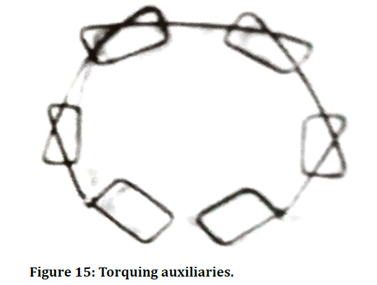
Figure 15: Torquing auxiliaries.
These auxiliaries and braking springs aid in anchorage of the anterior segment such that protraction of the molars can be carried out without further movement of anteriors.
Reverse labial root torque
The reverse labial root torque can be incorporated in the lower anteriors such that the roots of lower anteriors get engaged with the cortical bone, this aid in anchorage reinforcement. The reverse labial root torque can be achieved by two methods.
Torque in wire: The torque is incorporated in the archwire with respect to the anterior segment by twisting the arch wire in the anterior segment with the help of torquing pliers such that labial root torque is incorporated. This shifts the anchorage site from posterior to anterior allowing mesial movement of the molars (Figure 16).
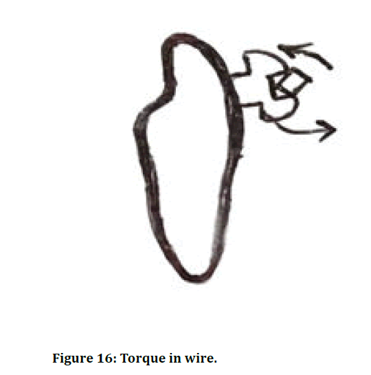
Figure 16: Torque in wire.
Brackets with negative torque: Brackets with negative torque can be bonded in the anteriors such that labial root torque is achieved (Figure 17).
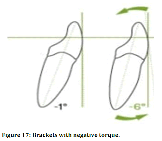
Figure 17: Brackets with negative torque.
En-mass retraction of mandibular posterior
Due to patient comfort issues, orthodontists generally do not prefer applying force system above the archwire level; hence the attention should wholly focus on forces of retraction. Therefore, the motto is a balanced combine archwire and bracket slot size for en-masse retraction of the molar. With the current scenario it can be easily said that the technology plays a very big role in dentistry, whether it be surgery or diagnosis or research. Recently a computed application has come into fame for its skills in determining the sites where optimum forces will be applied so as to gain good results for retraction. But due to the third molar impaction a whole lot of complication arise and one of them is alveolar bone resorption. Resorption in the bone causes difficulty in mesialization in the mandibular molar. Hence, it is very important to check the periodontal status of the patient before commencing the treatment of mesialization of the mandibular molar. Along with the help of technology calculation of the mesiodistal width of the molar is considered to be of great help in the treatment plan. A study was done on estimation of the mesiodistal width of the permanent mandibular molar in Sharad Pawar dental college. All these hardship and invention is to bring smile on the patients face. That one smile of satisfaction is worthall the efforts and all-nighters as facial appearance is one of the most important criteria for acceptance in the society and also for building one's self-confidence; hence, people who are satisfied with their appearance have more self-confidence.
Conclusion
The orthodontic force works on the level of the centre of resistance of the molar due to which the tooth is moved in the desired way and in the desired direction given good results. With the development of numerous techniques in orthodontics, mandibular molar mesialization or protraction is possible without much surgical intervention and therefore patient is more comfortable during orthodontic treatment. Molar protraction not only helps in treating edentulous space but also helps in normal eruption of the third molar. Since, with the usage of various appliances we can correct the edentulous space, prosthetic implants and bridges will no longer be needed in such patients and the treatment will be cost effective. Patient with malocclusion, open bite, excessive vertical growth pattern can be easily corrected with this technique. Hence this technique not only helps adults get rid of their edentulous space but also younger patients to correct their occlusion and facial profile. The department of orthodontics is different from other branches because it involves not only biology but also physics. There are various forces which move the tooth according to our will. This tells us how wonder the mechanism is of protraction; however even though this looks quite simple various theories and techniques make it complex. Many people think that the branch of orthodontics revolves around smile correction and facial profile correction but it is a misnomer. This branch not only provides esthetics to a patients smile and corrects the malocclusion but it also enables peaceful eruption of the third molar which is a huge achievement in itself.
References
- Siatkowski RE. Force system analysis of V-bend sliding mechanics. J Clin Orthod 1994; 28:539-546.
[Google Scholar] [PubMed]
- Kravitz ND, Jolley T. Mandibular molar protraction with temporary anchorage devices. J Clin Orthod 2008; 42:349-351.
[Google Scholar] [PubMed]
- Peretta R, Segu M. Cherry loop: A new loop to move the mandibular molar mesially. Prog Orthod 2001; 2:24-29.
- Vickers NJ. Animal communication: When i’m calling you, will you answer too. Curr Biol 2017; 27:13-15.
[Crossref] [Google Scholar] [PubMed]
- Burstone CJ, Koenig HA. Optimizing anterior and canine retraction. Am J Orthod 1976; 70:1-9.
[Crossref] [Google Scholar] [PubMed]
- Hoenigl KD, Freudenthaler J, Marcotte MR, et al. The centered T-loop-a new way of preactivation. Am J Orthod Dentofacial Orthop 1995; 108:149-153.
[Crossref] [Google Scholar] [PubMed]
- Yi J, Xiao J, Li Y, et al. Efficacy of piezocision on accelerating orthodontic tooth movement: A systematic review. Angle Orthod 2017; 87:491-498.
[Google Scholar] [PubMed]
- Dibart S, Keser EI. Piezocision™: Minimally invasive periodontally accelerated orthodontic tooth movement procedure. Compend Contin Educ Dent 2014; 14:119-44.
[Google Scholar] [PubMed]
- Al-Areqi MM, Abu Alhaija ES, Al-Maaitah EF. Effect of piezocision on mandibular second molar protraction. Angle Orthod 2020; 90:347-353.
[Google Scholar] [PubMed]
- Uribe F, Janakiraman N, Fattal AN, et al. Corticotomy-assisted molar protraction with the aid of temporary anchorage device. Angle Orthod 2013; 83:1083-1092.
[Crossref] [Google Scholar] [PubMed]
- Modi DN, Gupta DR, Borah DM. Newer orthodontic archwires-A review. Int J Appl Dent Sci 2020; 6:90-94.
- Li Y, Tang N, Xu Z, et al. Bidimensional techniques for stronger anterior torque control in extraction cases: A combined clinical and typodont study. Angle Orthod 2012; 82:715-722.
[Crossref] [Google Scholar] [PubMed]
- Freudenthaler JW, Bantleon HP, Haas R. Bicortical titanium screws for critical orthodontic anchorage in the mandible: A preliminary report on clinical applications. Clin Oral Implants Res 2001; 12:358-363.
[Google Scholar] [PubMed]
- Thote AM, Uddanwadiker RV, Sharma K, et al. Optimum force system for en-masse retraction of six maxillary anterior teeth in labial orthodontics. J Mech Med Biol 2020; 20:1950066.
- Thote AM, Patil RV, Shrivastava S. Estimation of orthodontic force parameters with developed computer application for en-masse retraction of six maxillary anterior teeth. Adv Prod Eng 2020; 497-505.
- Durge KJ, Bajaj P, Agrawal D. Distal molar surgery. J Evol Med Dent Sci 2020; 9:3335-3339.
- Tiwari MM, Jadhav VV, Daigavane P, et al. Meevik formula for estimating the mesiodistal width of permanent mandibular molar. J Evol Med Dent Sci 2021; 10:333-338.
- Verulkar A, Kamble R, Srivastav S, et al. Evaluation of the awareness of different orthodontic treatment appliances in patients undergoing orthodontic treatment of Maharashtra-A survey. Datta Meghe Inst Med Sci Univ 2020; 15:347-352.
- Kim SJ, Sung EH, Kim JW, et al. Mandibular molar protraction as an alternative treatment for edentulous spaces: Focus on changes in root length and alveolar bone height. J Am Dent Assoc 2015; 146:820-829.
[Crossref] [Google Scholar] [PubMed]
Author Info
Shambhavi S Moharil*, Kamble Ranjit and Renuka Talla
Department of Orthodontics, Datta Meghe Institute of Medical Sciences, Wardha, IndiaCitation: Shambhavi S Moharil, Kamble Ranjit, Renuka Talla, Mandibular Molar Protraction, J Res Med Dent Sci, 2023, 11 (08): 092-098.
Received: 15-Nov-2021, Manuscript No. JRMDS-23-47347; , Pre QC No. JRMDS-23-47347 (PQ); Editor assigned: 18-Nov-2021, Pre QC No. JRMDS-23-47347 (PQ); Reviewed: 02-Dec-2021, QC No. JRMDS-23-47347; Revised: 19-Jul-2023, Manuscript No. JRMDS-23-47347 (R); Published: 16-Aug-2023, DOI: https://www.longdom.org/admin/index.php?page=ft-preview&id=102712
