Research - (2022) Volume 10, Issue 5
Most Common Obturation Technique Used Between Anterior and Posterior Teeth
Abigail Ranasinghe and K Anjaneyalu*
*Correspondence: K Anjaneyalu, Department of Conservative and Endodontics, Saveetha Institute of Medical and Technical Science (SIMATS), Saveetha University, Chennai, India, Email:
Abstract
Introduction: Success of root canal therapy (RCT) is largely dependent upon the quality of biomechanical preparation and obturation of the pulp canal. A three dimensional seal of the root canal system is achieved by proper root canal obturation to prevent the recurrence of bacterial infection. The aim of this study is to determine the most common obturation technique used between anterior and posterior teeth.
Materials and methods: This study is a retrospective observational study conducted in a university hospital in Chennai. This study was carried out between the month of November 2020- March 2021.Data of patients who had undergone root canal treatment were included in the study sample. This was followed by Excel tabulation. Data was analysed using SPSS Software. The association of study variables was calculated using Chi Square test. Results: The age group between 20-30 years were the most common group which underwent root canal treatment within the given time period 27.9%. Most common obturation technique used was found to be matched tapered single cone technique 60.9% followed by cold lateral condensation technique 37.8%. Cold lateral condensation was predominantly used in anterior teeth whereas matched tapered single cone technique was predominantly used in posterior teeth. Conclusion: Cold lateral condensation technique is the gold standard technique and can be used in case of wider canals. Single tapered cone obturation technique has advantages over other techniques of obturation due to the fewer stress forces implied apically, thereby preventing an excess of sealer extrusion.
Keywords
Obturation, Anterior, Posterior, Cold lateral condensation, Single tapered cone
Introduction
Successful endodontic therapy is dependent on proper diagnosis, access cavity preparation, accurate determination of the working length, the thorough removal of microorganisms and their by-products through mechanical root canal instrumentation, antibacterial irrigation and adequate filling of the root canal space [1]. Proper endodontic diagnosis is an essential criterion in achieving successful treatment outcomes. In order to attain a good-quality obturation and a successful RCT, thorough cleaning and shaping of the root canal should be done to provide an ideal environment for the obturating material to fit and completely seal the pulp space [2]. A well-cleaned and -shaped root canal along with a three-dimensionally fitted pulpal space ensures the success of endodontic therapy [2]. If the root canal is not properly cleaned, shaped, or sterilized, regardless of the type of obturation method and obturating material, endodontic therapy cannot be successful [3]. Root canal disinfection involves removal of the bacterial biofilm that can be achieved by following a proper irrigation protocol, final irrigant activation and complete sealing of the root canal space [1]. Use of intracanal medicaments has also aided in disinfection of the root canal system, reducing inflammation.
Obturation is the filling and sealing of a prepared root canal with a root canal sealer and a core material. The core material occupies space while the sealer flows to areas of irregularities or those unaffected by mechanical preparation. An obturation must achieve a high level of adaptability to the prepared canal walls and the filling material must penetrate the dentinal tubules. The goal of root canal filling is to completely obliterate the canal space with a stable, nontoxic material and at the same time creating a hermetic seal to prevent the movement of tissue fluids, bacteria or bacterial by-products through the filled canal [4]. A three dimensional seal of the root canal system is achieved by proper root canal obturation to prevent the recurrence of bacterial infection. The microleakage between the root canal and the periapical tissues is hindered leading to death of any surviving microorganisms. This prevents the entry of nutrients and toxic bacterial products into the periapical tissues [5].
Various Obturation techniques used for filling the root canal system include lateral compaction, warm vertical compaction, and carrier-based obturation techniques. Cold Lateral compaction of GP is the gold standard technique [6]. This technique involves placing a single cone of gutta-percha (GP) with sealer in the prepared root canal and adding secondary GP cones that are compacted together with the use of a spreader. The cones stay together due to frictional grip and the presence of a sealer [7]. Although a time-consuming procedure, lateral condensation is preferred due to its low cost and controlled placement of GP in the canal [8]. The final mass is not homogenous and consists of numerous GP cones pressed together with the sealer filling most spaces in between. This technique of obturation is preferred in case of wide canals [9]. Disadvantages of lateral compaction technique to other techniques are that this technique of obturation cannot fill the root canal irregularities. In Carrier-based obturation technique, endodontic files are coated with thermoplasticized guttapercha [10]. Disadvantages of Lateral compaction and warm vertical compaction include lack of gutta-percha homogeneity, a high percentage of endodontic cement at the apical portion of the root and apical extrusion of gutta-percha. To overcome these disadvantages a single cone obturation technique was introduced.
The Single cone obturation technique is simpler than other techniques of obturation because of factors such as the operator is subjected to less fatigue and this technique of obturation is a more passive technique of obturation inducing lesser strain on the root canal system [11].
The single-cone technique of obturation uses a single cone with different tapers. This technique of obturation reduces the working time, allows faster filling, and is a less fatigue technique. This technique of obturation is similar to other techniques in terms of quality of the obturation, apical microleakage and bacterial penetration. Our team has extensive knowledge and research experience that has translate into high quality publications [12-31].
Materials and Methods
Study design
This study was a retrospective observational study conducted in a university setting. Approval for the project was obtained from the Institutional Review Board of Saveetha Institute of Medical and Technical Sciences, Chennai, India.
Sampling
10-fold serial dilutions for the collected saliva samples were prepared using sterile normal saline. Inoculate dilutions 10-3 on Sucrose- Bacitracin Agar (SB20) incubated first anaerobically for 48 hrs. at 37°C then aerobically incubated within 37°C for 24 hrs [8]. Data of patients were reviewed and then extracted. All patients who underwent endodontic treatment within 18-60 years in the given duration of time period were evaluated. Only relevant data was included to minimize sampling bias. Simple random sampling method was carried out. Cross verification of data for error was done by presence of additional reviewer and by photographic evaluation. Incomplete data collection was excluded from the study.
Data Collection
A single calibrated examiner evaluated the digital case records of patients who reported to Saveetha Dental College from June 2020- March 2021 and were reviewed. Inclusion criteria consisted of patients between the age group of 18-60 who underwent endodontic treatment. Exclusion criteria consisted of patients above 60 years, primary teeth, teeth with the presence of huge periapical lesions, severely calcified canals.
Statistical analysis
The collected data was tabulated and analysed with a statistical package for social sciences for windows, version 20.0 (SPSS Inc., Vancouver style) and results were obtained. Categorical variables were expressed in frequency and percentage. Chi square test was used to test association between categorical variables. Chi square test were carried out using age, gender and as independent variables and dependent variables. The statistical analysis was done by Pearson chi square test. P value < 0.005 was considered statistically significant.
Results
Based on the age of the patient, the age group evaluated for a maximum number of cases included 20-30 years, accounting 27.9% of overall cases,31-40 years accounted for 24.2% of overall cases, 41-50 years accounted for 21.5% of overall cases. 51-60 years accounted for 18.5 cases and 61-70 years age group accounted for 7.9% overall cases (Figure 1).
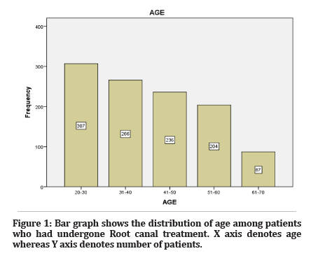
Figure 1: Bar graph shows the distribution of age among patients who had undergone Root canal treatment. X axis denotes age whereas Y axis denotes number of patients.
Based on the gender of the patient, males accounted for 46.3% cases, and females accounted for 53.7% cases (Figure 2).
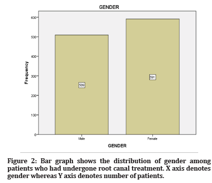
Figure 2: Bar graph shows the distribution of gender among patients who had undergone root canal treatment. X axis denotes gender whereas Y axis denotes number of patients.
Based on anterior/ posterior region anterior accounted for 45% cases and posterior accounted for 55% cases (Figure 3).
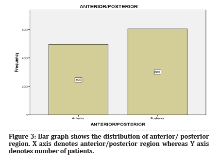
Figure 3: Bar graph shows the distribution of anterior/ posterior region. X axis denotes anterior/posterior region whereas Y axis denotes number of patients.
Based on the obturation technique matched tapered single cone technique accounted for 60.9% cases, cold lateral condensation technique accounted for 37.8% cases, warm lateral condensation accounted for 1% cases and thermafil obturation technique accounted for 0.3 cases (Figure 4).
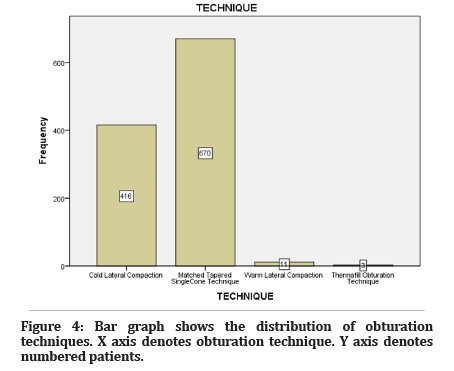
Figure 4: Bar graph shows the distribution of obturation techniques. X axis denotes obturation technique. Y axis denotes numbered patients.
Based on the site and obturation technique, maximum case in anteriors accounted for cold lateral condensation technique and in posteriors maximum cases accounted for matched tapered single cone technique (Figure 5).
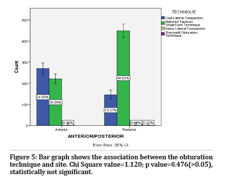
Figure 5: Bar graph shows the association between the obturation technique and site. Chi Square value=1.120; p value=0.476(>0.05), statistically not significant.
Discussion
In this study it is observed that an increased number of anterior root canal treatments were done when compared to posterior root canal treatment among dental students. Based on the tooth type, endodontic treatment of molars adds difficulties during root canal treatment for dental students with limited clinical experience. Tooth type was considered as a prognostic factor for root canal treatment according to several studies [32]. In this study it is seen that single cone obturation technique is widely used in posterior teeth when compared to other obturation techniques. Single cone technique of obturation is faster and easier in comparison with other techniques of obturation [33]. In terms of radiographic quality of obturation Lateral condensation is similar to single cone technique of obturation [34]. Single cone obturation systems, however, lacked a durable apical seal.
It is observed that in anteriors cold lateral compaction is predominantly used when compared to other techniques. Lateral compaction technique of obturation is the most commonly used technique and this technique of obturation is preferred in case of wide canals and has a durable apical seal [35]. In warm lateral condensation technique sufficient heating are essential in achieving a good adaptation in canals of widely varying diameter reported that multiple heating has shown to produce high levels of gutta-percha shrinkage [36].
Thermafil obturation technique which incorporates the use of thermal or frictional heat to plasticize the guttapercha, allowing for better adaptation to canal walls, higher degree of homogeneity and provide optimum apical and coronal sealing when compared to lateral condensation. However, this root canal obturation technique uses application of forces both laterally and apically to fill the canal in three-dimensionally and induce application of more stress on the root dentin. Single cone obturation technique is a passive obturation technique that prevents stresses on the root canal system and helps in maintaining the accurate placement of guttapercha and prevents extrusion of sealer. The use of a bidirectional spiral along with the single cone obturation technique helps to carry the sealer along the length, moving the excess cement coronally and providing a three-dimensional seal. The use of bioceramic sealers, along with single cone obturation technique, have proved to be beneficial.
This retrospective study was undertaken based on the obturation techniques used in permanent teeth to emphasise that achievement of three-dimensional obturation is an important aspect to prevent reinfection of the root canal system. The main purpose of endodontic treatment is to clean, shape and fill the root canal space thoroughly and prevent any interchange between the oral cavity, the root canal system and periradicular tissues, providing a barrier to reinfection. The success of endodontic treatment depends on adequate mechanical debridement of root canal and quality obturation with biocompatible material.
Conclusion
Within the limitations of the study it can be concluded that cold lateral condensation technique is the gold standard technique and can be used in case of wider canals. Single cone obturation technique was the most commonly used obturation technique and has several benefits such as the possibility of a faster endodontic treatment, less induction of apical forces thereby reducing the stress of the root dentin, prevention of excessive extrusion of the root canal sealer and gutta-percha.
Acknowledgement
The authors sincerely acknowledge the faculty Medical record department and Information technology department of SIMATS for their tireless help in sorting out datas pertinent to this study.
Funding
The present project is supported/funded/sponsored by Saveetha Institute of Medical and Technical Sciences, Saveetha Dental College and Hospitals, Saveetha University contributed by Anand Cycle Agency.
Conflict of Interest
None.
References
- Weis MV, Parashos P, Messer HH. Effect of obturation technique on sealer cement thickness and dentinal tubule penetration. Int Endod J 2004; 37:653-63.
- Rao R. Root Canal Morphology and Access Cavity Preparation. Advanced Endod 2009; 105.
- Ainley JE. Fluorometric assay of the apical seal of root canal fillings. Oral Surg Oral Med Oral Pathol 1970; 29:753-62.
- Manohar MP, Sharma S. A survey of the knowledge, attitude, and awareness about the principal choice of intracanal medicaments among the general dental practitioners and nonendodontic specialists. Indian J Dent Res 2018; 29:716.
- Bailey GC, Ng YL, Cunnington SA, et al. Root canal obturation by ultrasonic condensation of gutta‐percha. Part II: an in vitro investigation of the quality of obturation. Int Endod J 2004; 37:694-8.
- Gençoğlu N. Comparison of 6 different gutta-percha techniques (part II): Thermafil, JS Quick-Fill, Soft Core, Microseal, System B, and lateral condensation. Oral Surg Oral Med Oral Pathol Oral Radiol Endod 2003; 96:91-5.
- Levitan ME, Himel VT, Luckey JB. The effect of insertion rates on fill length and adaptation of a thermoplasticized gutta-percha technique. J Endod 2003; 29:505-8.
- Cueva-Goig R, Forner-Navarro L, Llena-Puy MC. Microscopic assessment of the sealing ability of three endodontic filling techniques. J Clin Exp Dent 2016; 8:e27.
- Cailleteau JG, Mullaney TP. Prevalence of teaching apical patency and various instrumentation and obturation techniques in United States dental schools. J Endod 1997; 23:394-6.
- Cueva-Goig R, Llena-Puy C, Forner-Navarro L. Apical sealing evaluation of three root canal filling techniques. Medicina Oral Patología Oral y Cirugia Bucal. 2012: S9–0.
- Collins J, Walker MP, Kulild J, et al. A Comparison of Three Gutta-Percha Obturation Techniques to Replicate Canal Irregularities. J Endod 2006; 32:762-5.
- Muthukrishnan L. Imminent antimicrobial bioink deploying cellulose, alginate, EPS and synthetic polymers for 3D bioprinting of tissue constructs. Carbohydr Polym 2021; 260:117774.
- PradeepKumar AR, Shemesh H, Nivedhitha MS, et al. Diagnosis of Vertical Root Fractures by Cone-beam Computed Tomography in Root-filled Teeth with Confirmation by Direct Visualization: A Systematic Review and Meta-Analysis. J Endod 2021; 47:1198-214.
- Chakraborty T, Jamal RF, Battineni G, et al. A Review of Prolonged Post-COVID-19 Symptoms and Their Implications on Dental Management. Int J Environ Res Public Health 2021;18.
- Muthukrishnan L. Nanotechnology for cleaner leather production: a review. Environ Chem Lett 2021; 19:2527-49.
- Teja KV, Ramesh S. Is a filled lateral canal - A sign of superiority? J Dent Sci 2020; 15:562-3.
- Narendran K, Jayalakshmi, Ms N, et al. Synthesis, characterization, free radical scavenging and cytotoxic activities of phenylvilangin, a substituted dimer of embelin. Indian J Pharm Sci 2020;82.
- Reddy P, Krithikadatta J, Srinivasan V, et al. Dental Caries Profile and Associated Risk Factors Among Adolescent School Children in an Urban South-Indian City. Oral Health Prev Dent 2020; 18:379-86.
- Sawant K, Pawar AM, Banga KS, et al. Dentinal Microcracks after Root Canal Instrumentation Using Instruments Manufactured with Different NiTi Alloys and the SAF System: A Systematic Review. NATO Adv Sci Inst Ser E Appl Sci 2021; 11:4984.
- Bhavikatti SK, Karobari MI, Zainuddin SLA, et al. Investigating the Antioxidant and Cytocompatibility of Mimusops elengi Linn Extract over Human Gingival Fibroblast Cells. Int J Environ Res Public Health 2021;18.
- Karobari MI, Basheer SN, Sayed FR, et al. An In Vitro Stereomicroscopic Evaluation of Bioactivity between Neo MTA Plus, Pro Root MTA, BIODENTINE & Glass Ionomer Cement Using Dye Penetration Method. Materials 2021; 14.
- Rohit Singh T, Ezhilarasan D. Ethanolic Extract of Lagerstroemia Speciosa (L.) Pers., Induces Apoptosis and Cell Cycle Arrest in HepG2 Cells. Nutr Cancer 2020; 72:146-56.
- Ezhilarasan D. MicroRNA interplay between hepatic stellate cell quiescence and activation. Eur J Pharmacol 2020; 885:173507.
- Romera A, Peredpaya S, Shparyk Y, et al. Bevacizumab biosimilar BEVZ92 versus reference bevacizumab in combination with FOLFOX or FOLFIRI as first-line treatment for metastatic colorectal cancer: a multicentre, open-label, randomised controlled trial. Lancet Gastroenterol Hepatol 2018; 3:845–55.
- Raj RK. β-Sitosterol-assisted silver nanoparticles activates Nrf2 and triggers mitochondrial apoptosis via oxidative stress in human hepatocellular cancer cell line. J Biomed Mater Res A 2020; 108:1899–908.
- Vijayashree Priyadharsini J. In silico validation of the non-antibiotic drugs acetaminophen and ibuprofen as antibacterial agents against red complex pathogens. J Periodontol 2019; 90:1441–8.
- Priyadharsini JV, Vijayashree Priyadharsini J, Smiline Girija AS, et al. In silico analysis of virulence genes in an emerging dental pathogen A. baumannii and related species. Arch Oral Biol 2018; 94:93-8.
- Uma Maheswari TN, Nivedhitha MS, Ramani P. Expression profile of salivary micro RNA-21 and 31 in oral potentially malignant disorders. Braz Oral Res 2020; 34:e002.
- Gudipaneni RK, Alam MK, Patil SR, et al. Measurement of the Maximum Occlusal Bite Force and its Relation to the Caries Spectrum of First Permanent Molars in Early Permanent Dentition. J Clin Pediatr Dent 2020; 44:423–8.
- Chaturvedula BB, Muthukrishnan A, Bhuvaraghan A, et al. Dens invaginatus: a review and orthodontic implications. Br Dent J 2021; 230:345–50.
- Kanniah P, Radhamani J, Chelliah P, et al. Green synthesis of multifaceted silver nanoparticles using the flower extract of Aerva lanata and evaluation of its biological and environmental applications. ChemistrySelect 2020; 5:2322–31.
- Benenati F, Khajotia S. A Radiographic Recall Evaluation of 894 Endodontic Cases Treated in a Dental School Setting. J Endod 2002; 28:391-5.
- Schäfer E, Nelius B, Bürklein S. A comparative evaluation of gutta-percha filled areas in curved root canals obturated with different techniques. Clin Oral Investig 2012; 16:225-30.
- Monticelli F, Sadek F, Schuster G, et al. Efficacy of Two Contemporary Single-cone Filling Techniques in Preventing Bacterial Leakage. J Endod 2007; 33:310-3.
- Hörsted-Bindslev P, Andersen MA, Jensen MF, et al. Quality of Molar Root Canal Fillings Performed With the Lateral Compaction and the Single-Cone Technique. J Endod 2007; 33:468-71.
- Venturi M, Breschi L. Evaluation of Apical Filling After Warm Vertical Gutta-Percha Compaction Using Different Procedures. J Endod 2004; 30:436–40.
Indexed at, Goggle Scholar, Cross Ref
Indexed at, Goggle Scholar, Cross Ref
Indexed at, Goggle Scholar, Cross Ref
Indexed at, Goggle Scholar, Cross Ref
Indexed at, Goggle Scholar, Cross Ref
Indexed at, Goggle Scholar, Cross Ref
Indexed at, Goggle Scholar, Cross Ref
Indexed at, Goggle Scholar, Cross Ref
Indexed at, Goggle Scholar, Cross Ref
Indexed at, Goggle Scholar, Cross Ref
Indexed at, Goggle Scholar, Cross Ref
Indexed at, Goggle Scholar, Cross Ref
Indexed at, Goggle Scholar, Cross Ref
Indexed at, Goggle Scholar, Cross Ref
Indexed at, Goggle Scholar, Cross Ref
Indexed at, Goggle Scholar, Cross Ref
Indexed at, Goggle Scholar, Cross Ref
Indexed at, Goggle Scholar, Cross Ref
Indexed at, Goggle Scholar, Cross Ref
Indexed at, Goggle Scholar, Cross Ref
Indexed at, Goggle Scholar, Cross Ref
Indexed at, Goggle Scholar, Cross Ref
Indexed at, Goggle Scholar, Cross Ref
Indexed at, Goggle Scholar, Cross Ref
Indexed at, Goggle Scholar, Cross Ref
Indexed at, Goggle Scholar, Cross Ref
Indexed at, Goggle Scholar, Cross Ref
Indexed at, Goggle Scholar, Cross Ref
Indexed at, Goggle Scholar, Cross Ref
Indexed at, Goggle Scholar, Cross Ref
Indexed at, Goggle Scholar, Cross Ref
Indexed at, Goggle Scholar, Cross Ref
Author Info
Abigail Ranasinghe and K Anjaneyalu*
Department of Conservative and Endodontics, Saveetha Institute of Medical and Technical Science (SIMATS), Saveetha University, Chennai, IndiaCitation: Abigail Ranasinghe, Subash Sharma, Most Common Obturation Technique Used Between Anterior and Posterior Teeth, J Res Med Dent Sci, 2022, 10 (5):56-60.
Received: 25-Apr-2022, Manuscript No. JRMDS-22-60230; , Pre QC No. JRMDS-22-60230 (PQ); Editor assigned: 27-Apr-2022, Pre QC No. JRMDS-22-60230 (PQ); Reviewed: 10-May-2022, QC No. JRMDS-22-60230; Revised: 15-May-2022, Manuscript No. JRMDS-22-60230 (R); Published: 22-May-2022
