Research - (2021) Volume 9, Issue 4
Oral Manifestations a Key in Diagnosing Iron Deficiency AnemiaâA Case Report
Saraswathi Gopal K, Srividhya S and Sushmitha S*
*Correspondence: Sushmitha S, Department of Oral medicine and radiology, Meenakshi Academy of Higher Education and Research, Faculty of Dentistry, Meenakshi Ammal Dental college, India, Email:
Abstract
Iron deficiency anaemia is one of the most common nutritional deficiencies disorder worldwide. It is characterized by symptoms like fatigue, weakness, pallor and koilonychias. Oral mucosa can reflect the general status of the patient and these signs can sometimes precede general symptoms. Oral mucosal pigmentations and depapillationof tonguecan also be an initial presentation in iron deficiencies anaemia. Thus, dentists play an important role in early diagnosis of anemia enabling a prompt referral in certain cases. With this background we report a case of iron deficiency anaemia with oral manifestations.
Keywords
Anaemia, Iron deficiency anaemia, Koilonychia, Haemoglobin, Oral manifestations, Angular chelitis
Introduction
A lack of significant iron in the normal red blood cells leads to iron deficiency anemia. It is the most common type of anemia especially in children and women [1]. Prelatent, latent and the marked iron deficiency are the three well known stages of iron deficiency anemia [2]. A variety of underlying causes of an inadequate iron supply oran increased iron requirements could be a possible etiological factor. Recovered iron from the senescent erythrocytes are said to produce most of the Iron for erythropoiesis and only 1-2 mg are absorbed from the diet [3]. Hepcidin a systemic hormone that is produced in the liver is the key hormone that inhibits iron absorption from the intestine as well as its release from macrophage stores.
Clinically in a case of iron-deficiency anemia it is said that the storage iron declines until iron delivery to the bone marrow is insufficient for erythropoiesis. The clinical indicators such as low plasma ferritin, followed by decrease in plasma iron - transferrin saturation culminating with low-hemoglobin content in red blood cells are generally monitored [4]. Iron deficiency symptoms are generally secondary to anaemia and include symptoms like weakness, headache, fatigue, koilonychia spoon shaped nails and exercise intolerance. Oral manifestations of anaemia are well recognised that includesglossitis, glossodynia, pallor of oral mucosa, angular cheilitis, erythematous mucositisetc [5]. A basic change in the metabolism of oral epithelial cells is said to play a key role in the initiation of oral manifestation in these cases [6]. Here in this article, we present a case of Iron deficiency anaemia in a 45-year-old female with oral manifestations.
Case Report
A 45-year-old female patient reported to our department with the chief complaint of dryness and bleeding from the corner of her lip for the past 4 months. Patient gave a history of mild pain, intermittent type in relation to the corner of the lips that aggravated on chewing with difficulty on mouth opening. History of burning sensation that aggravated during food intake for the past few months. Patient also gave a history of brown discoloration noted in her inner cheeks few months back. Medical History revealed history of Menorrhagia for the past 2 years.
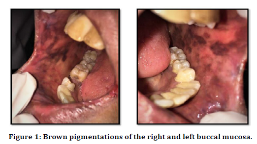
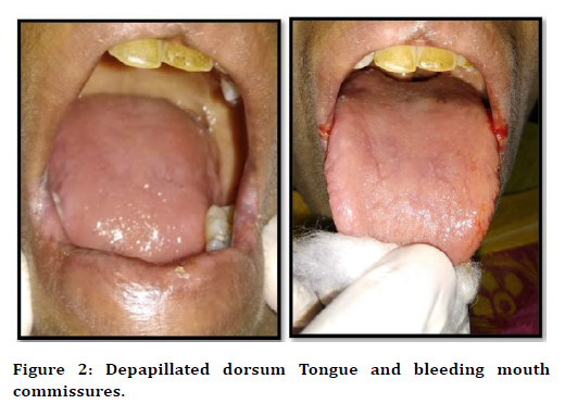
On General examination a positive finding of dry skin, pallor brittle hair, paler lower palpebral conjunctiva and nail bed were noticed. On intraoral inspection of the right and left buccal mucosa generalized pale mucosa was evident intermixed with diffuse brown hyper pigmented areas (Figure 1). The dorsum tongue appeared paler with generalized depapillation and glossiness (Figure 2).

Figure 1. Brown pigmentations of the right and left buccal mucosa.

Figure 2. Depapillated dorsum Tongue and bleeding mouth commissures.
The skin folds at the right and left lip commissures appeared inflamed, hyperkeratotic with mild cracks. On palpation the pigmented lesion in relation to the right and left buccal mucosa appeared to be non-tender. The lesion at the right and left lip commissures appeared tender on palpation with bleeding evident on palpation. Correlating with the clinical findings and patient’s history a provisional diagnosis of Anaemic stomatitis with angular chelitis was given. Further the patient was subjected for complete haemogram analysis which revealed the following values (Table 1) and the peripheral smear showed RBC’s appearing as microcytic hypochromic cells with Moderate Anisopoiklocytosis, tear drop and pencil cells. An Impression suggestive of Microcytic Hypochromic Anemia was given. Hence a final diagnosis of Anemic stomatitis & angular cheilitis due to iron deficiency anaemia was given correlating clinical presentation and blood investigations. Further the patient was referred to general physician and was under pharmacological management.
| RBC count | 2.1 millions/cubic mm |
|---|---|
| Hb% | 6.2% gm/dl |
| WBC count | 4200 cells/c.mm |
| Platelets | 4.0 lakhs /c.mm |
| Serum Iron | 10.9 microgram/Dl |
| Total iron binding capacity | 512.6 microgram/Dl |
Table 1: Haemogram analysis.
Discussion
Anaemia is defined as reduction in the oxygen carrying capacity of blood, due to reduction in number of circulating RBCs or abnormality in Hb within Red blood cells. According to the WHO criteria when the Hb level <13g/dL in men and<12g/dL in women it is anemic [6]. Globally, anaemia is said to affect 1.62 billion people that corresponds to nearly 24.8% of the population. The highest prevalence is seen in preschoolage children in 47.4% cases and the lowest prevalence is in men 12.7% [7]. Identifying anemia is especially important as it is a debilitating condition that can induce or worsen an existing disease like congestive heart failure and can cause cognitive impairment, dizziness and apathy impairing quality of life [8].
Iron deficiency is one of the main etiologic factors of anemia. It may cause because of three main etiological factors like an Increased demand for Iron, Increased iron loss or may be due to decreased Iron absorption. The main etiological cause of this deficiency in a huge population is thought due to Low dietary intake like iron, folic acid, and vitamin B12, and other factors being underlying chronic diseases and chronic blood loss due to hookworm infestation and malaria [9].
The most abundant iron containing protein in the human body is haemoglobin which accounts for nearly one half of the total iron in the body. Erythropoiesis-related demands for iron are created by three variables: tissue oxygenation, erythrocyte turnover, and erythrocyte loss from hemorrhage. About 20 ml of senescent erythrocytes are eliminated daily and from that about 20 mg of iron are being recycled. So when there is a hemorrhage additional iron must be absorbed from the diet to meet the steady-state demands of the host.If iron losses continue, the newly produced erythrocytes will have decreased hemoglobin, causing the amount of iron provided by the same number of senescent erythrocytes to be reduced [7]. The bone marrow tries to pump out all the RBC’s before maturing to meet the demand, thus resulting in altered shape –Poikilocytosis and altered shape – Anisocytosis of the cells.
The oral signs and symptoms of anemia are glossitis, glossodynia, angular cheilitis, recurrent oral ulcer, oral candidiasis, erythematous mucositis, and palor of oral mucosa. In some cases, the initial manifestation of anemia of chronic diseases may present in the oral cavity beforethe development of general symptoms. The changes in the oral cavity are thought to be due to abnormalities in the cell structure and the keratinization pattern of the oral epithelium [10].
The general signs and symptoms such as pallor, fatigue and dyspnea may be overlooked in elderly as these findings may be attributed to old age. The other systemic sign of anemia is the Knuckle pigmentation, Brittle nails, Koilonychia, Hair loss, Restless leg syndrome etc. The main pathogenesis behind the formation of spoon shaped nails is because of decreased in the subungual blood supply in the nail bed creating a dip in the nail bed. The other pathogenesis for the manifestation of Brittle nails is hypothesized as due to the loss of cysteine disulfide bonds because of lack of oxygen which aids in giving a hard texture to the nail bed. Thus, the oral signs and symptoms in these cases offers the dentist a key role in earlier identification of this underlying debliating condition.
On biochemical analysis there will be a reduction in RBC count, serum Iron, serum Ferritin, Transferrin saturation and hepcidin level. The Total iron binding capacity will be increased along with the Serum Levels of Transferrin Receptor Protein [7]. All these parameters will aid in the diagnosis of this condition. The management of these conditions are done through blood transfusions especially in the cases of acute blood loss. An oral iron therapy of 200 mg is generally prescribed for 6- 12 months where it is said that only 50 mg per day of iron absorption takes place. In cases where patient is intolerable to oral iron therapy a parenteral route of iron therapy is prescribed. The main complication in this route is the anaphylaxis [11-13].
Conclusion
Thus, Mouth is referred to as the mirror of the body, the oral mucosa reflects the general health status of a person. These changes detected on the tongue and labial mucosa in certain anaemic cases may precede general systemic features. Thus, dentist during routing oral screening can aid in identifying these clinical features thereby providing an early diagnosis and prompt treatment with referral to the physician thereby improving the quality of life of patients.
References
- Johnson-Wimbley TD, Graham DY. Diagnosis and management of iron deficiency anemia in the 21st century. Therapeutic Adv Gastroenterol 2011; 4:177-184.
- Ozdemir N. Iron deficiency anaemia from diagnosis to treatment in children. Turk Ped Ars 2015; 50:11-19.
- Handelman GJ, Levin NW. Iron and anemia in human biology: a review of mechanisms. Heart Fail Rev 2008; 13:393-404.
- Andrews NC, Schmidt PJ. Iron homeostasis. Annu Rev Physiol 2007; 69:69-85.
- Torsvik IK, Markestad T, Ueland PM, et al. Evaluating iron status and the risk of anaemia in young infant using erythrocyte parameters. Int Paediatr Re Foundation 2012; 73:214- 220.
- Adeyemo TA, Adeyemo WL, Adediran A. et al. Orofacial manifestations of hematological disorders: Anemia and hemostatic disorders. Indian J Dent Res 2011; 22:454.
- Rammohan A, Awofeso N, Robitaille MC. Addressing female iron-deficiency anaemia in India: Is vegetarianism the major obstacle? ISRN Public Health 2012; 8.
- Kawaljit Kaur. Anaemia ‘a silent killer’ among women in India: Present scenario. Eur J Zoological Res 2014; 3:32-36.
- Pontes HA, Neto NC, Ferreira KB. et al. Oral manifestations of vitamin B12 deficiency: A case report. J Can Dent Assoc 2009; 75:533-537.
- Raju V, Arora A, Saddu S. Atrophic glossitis: An indicator of iron deficiency anemia: Report of three cases. Int J Dent Clin 2014; 6:30-31.
- DeRossi SS, Raghavendra S. Anemia. Oral Surg Oral Med Oral Pathol Oral Radiol Endodontol 2003; 95:131-141.
- Wu YC, Wang YP, Chang JYF, et al. oral manifestations, and blood profile in patients with iron deficiency anemia. J Formosan Med Assoc 2014; 113:83–87.
- Darby WJ. The oral manifestations of iron deficiency. J Am Med Assoc 1946; 130:830.
Author Info
Saraswathi Gopal K, Srividhya S and Sushmitha S*
Department of Oral medicine and radiology, Meenakshi Academy of Higher Education and Research, Faculty of Dentistry, Meenakshi Ammal Dental college, Chennai, Tamil Nadu, IndiaCitation: Saraswathi Gopal. K, Srividhya.S, Sushmitha.S, Oral Manifestations a Key in Diagnosing Iron Deficiency Anemiaâ??A Case Report, J Res Med Dent Sci, 2021, 9 (4): 361-364.
Received: 20-Mar-2021 Accepted: 08-Apr-2021
