Research - (2021) Volume 9, Issue 7
The Influence of Gonial Angle on the Incidence of Mandibular Angle Fracture and Evaluation of Treatment Modalities
Dalya Abbood1* and Waleed Khalil Ismael2
*Correspondence: Specialist oral and maxillofacial surgeo. Dalya Abbood, Al-Kindy teaching hospital, Iraq, Email:
Abstract
Background: Fractures of the mandible are the most common facial fractures, despite considerable collective experience and extensive literature on the subject; some aspects of care still remain controversial. Anatomically there is several regional classifications for fracture sites. Each fracture site has different etiological complex factors according to the force, direction, and the nature of trauma also there are endogenous factors may be had relation with fractures evolving mechanism. This study discusses one factor about the gonial angle measurements and its relation to the fracture site, management work up and possible treatment options. Aims of study: The aims of this prospective study are to analyse the association between the gonial angle measurement and the incidence of mandibular angle fracture radiographically and to evaluate the outcome of different treatment modalities clinically and radiographically. Materials and Methods: Between January 2017 and August 2018, in Al-Yarmuk teaching hospital, the maxillofacial department, (97) patients with fracture mandible were admitted, (36) patients from them had angle fracture. The data for (61) patients were used to compare with included data (Digital precise method were used to measure the mandibular gonial measurement parameter), all data were collected carefully and full history and examination achieved with selective radiological investigation (othropantography) were chosen according to patient data sheet and the outcomes of different treatment modalities were evaluated by using appropriate analytic and statistical Methods and software for representation of the data results. Results: The findings about the relation of the gonial angle measurements to the mandibular angle fracture were significantly remarkable, the mean measurement of gonial angle between patients with angle fracture were (128.3 ± 12.5) which was higher than others non-angled fractures also this result were tested and (P value=0.00728, also impacted 3rd molar tooth among patients is also included and the results was significant, and the high gonial angle measurements (mean=137.6) was significantly positive for selection of the type of managements (open reduction), (p value=0.0359). Conclusions: High gonial angle in early orhtopantography at admission time may be predicted for probable mandibular angle fractures more than others fracture sites also higher angle measures may be predictive factor for advance open reduction surgery. The impacted 3rd molar tooth may be acted as aggravating factors for developing angle fractures.
Keywords
Gonial angle, Mandibular fracture, Impacted 3rd molar, OPG
Introduction
The incidence of mandibular fracture is more commonly than other bony fractures of the facial skeleton, which could be related to its prominent position and exposed situation.
The strength of the mandible is determined by various factors such as the presence of active and strong musculature, the shape and thickness of bone, and the presence or absence of teeth [1]. Mandibular angle fractures follow a pattern common to many injuries and this depends on multiple factors including direction, amount of force, presence of soft tissue bulk, and biomechanical characteristics of the mandible such as bone density, mass or anatomic structures creating weak areas [2].
Fractures of the mandibular angle are common in occurrence. The higher Incidence has been attributed to the curvature at the angle region, the presence of impacted third molars, and the height of the mandible at the angle. The poor quality of bone at the angle region also has been described as a cause of fracture [3].
One important anthropometric feature that describes the mandibular growth pattern is the mandibular gonial angle. It refers to the angle that is formed by the ramus line and the mandibular line, where the ramus line is tangent to the posterior border of the mandible and the mandibular line is the lower border of the mandible through the gnathion [4].
The gonial angle can be assessed clinically or radiographically by manual and digital methods. In an individual, although the right and left gonial angles are the same, the normal gonial angle varies according to ethnicity, gender, and age. Based on the gonial angle, individuals can be classified as having a high, normal, or low angle or a vertical, normal, or horizontal growth, respectively. Numerous studies have established a positive correlation between the gonial angle and the bony architecture at the mandibular angle region. Further, a high gonial angle has been associated with weaker bite forces and decreased cortical thickness [5].
The presence of third molar is associated with twofold to threefold increased risk of angle fractures compared with the absence, and the fractures are most likely to occur in teens and those in their twenties.
This is of clinical interest because this age is most likely to have unerupted third molar [6,7]. The presence of third molars has been suggested to contribute to an increased mandibular fragility because the mandible loses part of its bone structure to harbour tissues that do not contribute to its strength [8]. There is a retrospective study shows overwhelming evidence of a direct relation of impacted third molars to increased incidence of angle fractures. The absence of impacted third molars is directly related to shift in fracture incidence from angle to the condylar region [9]. The modalities for the treatment of fractures of the mandible have been in a constant state of evolution. Fractures of the angle of the mandible are technically challenging and many techniques for treatment of these fractures have been proposed in literature. Despite numerous advances angle fractures remain amongst the most difficult and unpredictable to treat as compared with those of other areas of mandible. Fractures of the mandibular angle are plagued with the highest rate of complications amongst all mandibular fractures [10].
Materials and Methods
An observational prospective study conducted in Al- Yarmuk Teaching Hospital from January 2017 to August 2018, for patients presented with fracture mandible due to any cause of trauma.
From a total No. of (97) patients with fractured mandibles there were (36) patients confirmed as angle fractures, which represented as (31) males (86.11%) and (5) females (13.89%) aged ranged between 18 - 60 years (mean =29.66) that fulfilled the inclusion criteria were participated in this study.
All the cases diagnosis was approved by clinical examination and OPG.
Inclusion criteria
The inclusion criteria for sample selection consisted of preoperative digital OPGs for patients.
Exclusion criteria
- OPGs showing a completely edentulous state.
- Pathological changes, such as cystic lesions and osteoporosis.
- Patients with pan facial fracture.
- Patients with mandibular asymmetry.
- Patients with bilateral angle fracture.
Diagnosis of mandibular fractures
Diagnosis was based on clinical and radiographical examination:
Clinical examination
Extra-oral: The patients were examined by inspection for any facial asymmetry, ecchymosis, swelling, soft tissue laceration, obvious deformity of bony contour, and by palpation for the presence of tenderness over the fracture site, step defect or bony crepitus, anaesthesia or paraesthesia of lower lip and limitation of mouth opening.
Intra-oral: Included examination f o r any ecchymosis or hematoma especially in the lingual sulcus which is the pathognomonic feature of a fracture, any step defect in occlusion or alveolus and obvious laceration in the overlying mucosa, occlusal disturbance, tenderness and step deformities in the bone.
Radiographical examination
A pre-operative radiographical examination was obtained for all patients in order to diagnose and assess the site and extent of the fractures. The views included:
Orthopantomogram (OPG)
Fracture site and side.
The presence of associated fractures.
The presence of impacted 3rd molar and its class.
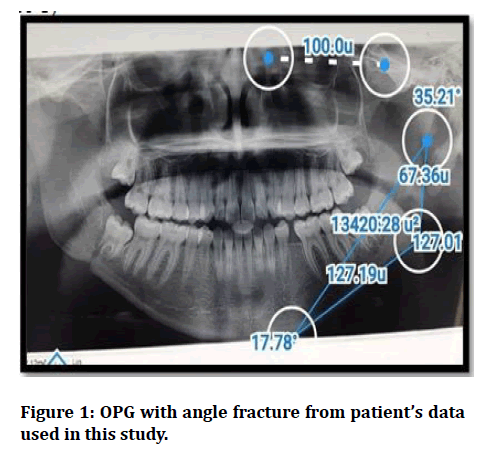
Measuring of Gonial angle (The 3-point angular measurement of the gonial angle was determined by digitally calculating the angular measurement formed by the points connecting the articulare, the gonion, and the menton.
The normal range for the gonial angle was fixed at (121.8 ± 6.2) based on norms specific to the present study population. Any value larger (128) was considered a high gonial angle and any value smaller than (115.6) was considered a low gonial angle (Figure 1).

Figure 1: OPG with angle fracture from patient’s data used in this study.
Lines of treatment
The surgical preparations begins with draping of the skin with 10% povidone Iodine and the oral cavity with chlorhexidine followed by copious irrigation with normal saline solution and patients were draped in the usual manner. There were three treatment modalities of fractured mandible, which were conservative, closed reduction and open reduction.
Conservative
When the fracture was undisplaced it was been treated conservatively by keeping the patient on soft diet for one month.
Closed reduction
Reduction of the fracture manually, in case the arch bar (Erich type) was used; it was inserted in the upper and lower jaw.
A suitable length of arch bar cut and bent to correct shape before operation, then 15 cm length of stainless wires (0.45 or 0.35) were stretched to 10% from this length to avoid slacking, then the teeth tied on the bar by twisting the wires.
When eyelet wiring is used, holding 15 cm length of wire by pair of artery forceps and gave the middle of wire to turns around a piece of 3 mm in diameter, then the eyelets were fitted between two teeth.
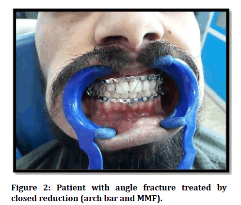
Then the tie wires inserted between upper and lower arch bars or eyelets for immobilization for 21 days for healthy young patient added one week if the patient is older than 40, smoker, has multiple unilateral fractures, or presented tooth in fracture line (Figure 2).

Figure 2: Patient with angle fracture treated by closed reduction (arch bar and MMF).
Open reduction
The access to the fracture line is either
Intraorally
By vestibular incision about 3 cm distal to the 2nd premolar extended to the external oblique ridge until the ascending ramus, the mucoperiosteal flap was elevated to expose lower border of mandible and the fracture site. After reducing the fracture manually IMF was placed to the arch bar or eyelet.
The fracture was fixed by mini plate; it was positioned according to Champy technique. The plate held to the bone surface while drilling the screw holes monocortically under irrigation.
When transosseous wiring was used, in cases of upper border wiring, holes were drilled in the bone ends on each side of fracture line then suitable length of 0.5 mm stainless-steel wire is passed through the holes, after reduction, the two ends of wire were twisted. Then the wound is closured.
Extra orally
In this study submandibular approach (Risdon approach) was used and the fracture line is also access extra orally. After marking the inferior border and angle of the mandible, the skin incision was done about two cm below the inferior border of the mandible. The incision is carried down through the skin and subcutaneous tissue to the level of the platysma muscle which is divided to expose the superficial layer of the deep cervical fascia which is divided and the dissection continues bluntly under the fascia to the inferior border of the mandible to protect the marginal mandibular branch of the facial nerve. The facial vessels when encountered and it was ligated then the incision extends more posteriorly to expose the angle region, the periosteum is incised and the mandible exposed. After exposure of the fracture it reduced manually MMF is placed and then fixation by titanium reconstruction plate, it was positioned at the inferior border of the mandible after bent using pliers and benders so it placed passively on the bone surface. The plate was held to the bone surface while drilling the holes through bicortical under irrigation with normal saline. The appropriate fixation was done by screws in holes. Then the wound closure was done in layers without tension.
Postoperative care and follow-up
All the patients were given intravenous systemic antibiotic postoperatively. The follow up of patients were done every week for one month postoperatively and then once monthly in the subsequent months for a period of 3 months . In the close reduction treatment IMF released after 21 days, while in open reduction treatment IMF released after 2 weeks. Successful treatment was regarded as stable bone, gaining the pre-trauma occlusion, absence of clinical infection and pain at the fracture site during function. Complications ( early complication like infection, pain, found dehiscence, plate exposure) was conditions arising in patients and these were occurred during nd after treatment but probably persisted beyond eight weeks from the commencement of treatment (late complications).During the follow up, postoperative complications were assessed, recorded and managed.
Statistical analysis
The statistical analysis was performed using SPSS windows version 23 Software and also Excel sheet used for collecting the data information’s .Suitable quantitive analytics models used for comparing of the data and compiling the result then the tables and graphs were used to describe this results. Chi’s square test were used to test qualitative and frequency data. P value ˂ 0.05 was considered significant.
Results
According to the data of this study, the most common age group for mandibular angle fractures was (20-29 years) with (36.11%). The mean was (29.66 years), also the sex distribution was (31) male (86.11%) and (5) female (13.89%). In this study and according to the arrangement of the occupations groups the results were (19.44% for employed (qualified), 41.67% for employed (nonqualified), 27.78% for students/post graduated group, and 11.11% for non- employed.
For alcoholic consumption group according to this study result were (44.4%) for positive and (55.6%) for negative consumption parameter. For the causative factors of trauma were presented as (Assault 27.78%, RTA 33.33%, MVA 16.67% (total was 50%), Fall from height 13.89%, sport related injuries 8.33%) (Table 1).
Table 1: Comparative table among trauma causative factors.
| Cause of trauma | No. | % |
|---|---|---|
| Assault | 10 | 27.78% |
| RTA | 12 | 33.33% |
| FFH | 5 | 13.89% |
| Sport | 3 | 8.33% |
| MVA | 6 | 16.67% |
| Total | 36 | 100.00% |
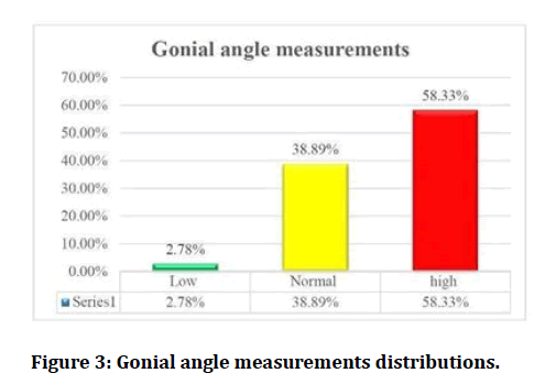
For restraining availability factor according to this study was (93.33% for non-restrained drivers and only 6.67% for restrained), these data were represented (18) patients with RTA or MVA. The total No. of patients in this study were (97) but there were only (36) patients with approved angle fracture while (61) were non-angle fracture with associated other fractures and among those (61) patients there were multiple fractures which totally equal (101). And from the patients with angle fracture were (20) patients of them were came with another fractures in the mandible (associated fracture), and were presented as body 19.44%), Para symphysis and symphysis (36.11%) . and (16) patients of them had isolated angle fracture (not associated with other fractures) (44.45%). (Table 2) In this study the gonial angle measures were classified to low, normal and high categories, and the results were Low (2.78%), normal (38.89%), high (58.33%) (Figure 3).
Table 2: Comparative table between angled and non-angled fractures and their relation to gonial angle measurements.
| Site of fractures | No. of Pts | No. of # | % of # | gonial angle mean ± SD |
|---|---|---|---|---|
| Angle fracture | 36 | 36 | 26.27 | 128.3±12.5 |
| Non angle fracture | 61 | |||
| Condyle | 3 | 24.09 | 124.1±7.1 | |
| Body | 2 | 17.52 | 122.5±4,8 | |
| symphysis and parasymph -ysis | 44 | 32.12% | 122.8±4.1 | |
| Total | 97 | 137 | 100 | |
| P-Value | 0.00728* | |||

Figure 3: Gonial angle measurements distributions.
For impacted 3rd molar relation with mandibular angle fracture, Total number of impacted 3rd molar were (9) cases (25%) and by applying Winter’s classification Mesio-angular was (44.5%), Vertical (33.3%), Horizontal (22.2%) and disto-angular and others rare classification (0%). Also when we applyed Pell and Gregory’s classification in measurement of 3rd molar impaction and the results were for Vertical position (related to occlusion plane) A cl (33.3%) , B cl (55.6%) , C cl (11.1%) .and for Horizontal position (related to anterior border of the ramus) I cl (55.6%) , II cl (33.3%) , III cl (11.1%) (CL :classification).
The management protocol classified into 3 options and our study result represented as conservative (8.33%), Closed reduction (72.22%), Open reduction (19.4%). The relation of management types between the mean of gonial angle measures for the patients and are for conservative (122.5), close reduction (126.7) and open reduction (137.6). In this study the results for postoperative complications were early complication during IMF treatment: Infection (8.30%), Pain on Fracture site (0%), Wound dehiscence (2.80%), Plate exposure (2.80%)
Late complication after removal IMF
Occlusal disturbance (8.30%), Neural Deficit (30.60%), limited mouth opening ( 19.40%), Non-union (0%), Fibrous union (0%), mal-union (0%), Satisfiction of patient (16.67%).
Discussion
According to this study the Age, sex, causative factors, alcohol consumption, and restraining availability are in similar value to worldwide standards in others studies used [11-14] respectively, which are significantly related to male sex, young age group, road traffic accident, consumption of alcohol during driving and trauma severity will be reduced when there is restraining equipment used in the vehicles [11].
This study uses statistical analysis and table to compare between site of the fracture (angled and non-angled) and enumeration of the number of each fractures and percentage also the gonial angle measurements mean were compared in statistics analysis and p-value calculated which was significant for the result of proportion of high angle measurement with angle site of mandibular fractures.
The presence of specific types of Impacted 3rd molar according to winter’s or Pell and Gregory’s classifications may have a role in developing that fracture more than other types. The presence of the third molars resulted in a difference in the stress distribution. Also found that it was noticeable that the impact of force on the chin resulted in a concentration of stress on the external oblique ridge, and when the third molar was present, this concentration extended to the alveolar process and on the vestibular aspect of the mandibular angle when the third molar was present, and on the condylar neck when it was absent [12].
The closed reduction is more frequent chosen method of treatment in this study because of the most common fractures is favourable fractures and also open reduction required more facilities, cost- benefit and unavailability of required equipment. The conservative option is preserved for no displaced fractures which is few cases in this study [13].
The relation of management types between the mean of gonial angle measures for the patients and are for conservative (122.5) , closed reduction (126.7) and open reduction (137.6).
And there is No other comparative study mentioned this hypothesis about the relation of the angle measurement with choosing the type of managements. The main hypothesis presented by this study is the higher probability of choosing open reduction management when there is a preoperative higher gonial angle measurement. When the study is compared to other comparative studies, this study result were better outcome than other study in many categories of the postoperative complications and this finding may be due to high rate of surgical managements in the comparative study (75% of cases) when we compare to our study (19.4%) [14].
Conclusion
The high measurements of gonial angle are highly associated with angle fractures (fact) and more. Higher measurements are directly proportion to increase the probability of more advance surgical procedures (Open reduction). Prescience of Impacted 3rd molar tooth is probably predisposing factor for developing angle fractures. Higher incidence between non- qualified employees and alcohol consumers. RTA/MVA are represented the most causative facial trauma. High incidence of assault accidents victims in Iraq more than other studies. High percentage of non-restrained drivers in our country.
References
- Banks P, Brown A. Fractures of the Facial Skeleton. StLouis, MO, Butterworth-Heinemann, 2005; 6.
- Lee JT, Dodson TB. The effect of mandibular third molar presence and position on the risk of an angle fracture. J Oral Maxillofac Surg 2002; 58:394-9.
- Panneerselvam E, Prasad PJ, Balasubramaniam S. The influence of the mandibular gonial angle on the incidence of mandibular angle fracture-A radiomorphometric study. J Oral Maxillofac Surg 2017; 75:153-159.
- Upadhyay RB, Upadhyay J, Agrawal P, et al. Analysis of gonial angle in relation to age, gender, and dentition status by radiological and anthropometric methods. J Forensic Dent Sci 2012; 4:29-33.
- Metin M, Sener I, Tek M. Impacted teeth and mandibular fractures. Eur J Dent 2007; 1:18-20.
- Meisami T, Sojat A, Sàndor GK, et al. Impacted third molars and risk of angle fracture, Int J Oral Maxillofac Surg 2002; 31:140-4,2002
- Menon S, Kumar V, Srihari V, et al. Correlation of third molar status with incidence of condylar and angle fractures. Craniomaxillofac Trauma Reconstruction 2016; 9:224-8.
- Ellis E, Walker L. Treatment of mandibular angle fractures using one noncompression miniplate. J Oral Maxillofac Surg 1996; 54:864–871.
- arolina Larrazabal-Moron, Juan A. Sanchis-gimeno: Gonial angle growth patterns according to age and gender annals of anatomy. Anatomischer Anzeiger 2018; 215: 93-96.
- Subbaiah MK, Ponnuswamy IA, David MP. Relationship between mandibular angle fracture and state of eruption of mandibular third molar: A digital radiographic study. J Indian Academy Oral Med Radiol 2015; 27:35.
- Bataineh AB. Etiology and incidence of maxillofacial fractures in the north of Jordan. Oral Surg Oral Med Oral Pathol Oral Radiol Endodontol 1998; 86:31-5.
- Anyanechi CE, Saheeb BD. Complications of mandibular fracture: Study of the treatment methods in Calabar, Nigeria. West Indian Med J 2014; 63:349.
- Gadicherla S, Sasikumar P, Gill SS, et al. Mandibular fractures and associated factors at a tertiary care hospital. Arch Trauma Res 2016; 5.
- Iida S, Kogo M, Sugiura T, et al. Retrospective analysis of 1502 patients with facial fractures. Int J Oral Maxillofac Surg 2001; 30:286-90.
Author Info
Dalya Abbood1* and Waleed Khalil Ismael2
1Al-Kindy teaching hospital, Iraq2Department of Maxillofacial, Al- Yarmuk teaching hospital, Iraq
Citation: Dalya Abbood, Waleed Khalil Ismael,The Influence of Gonial Angle on the Incidence of Mandibular Angle Fracture and Evaluation of Treatment Modalities, J Res Med Dent Sci, 2021, 9(7): 251-256
Received: 26-May-2021 Accepted: 07-Dec-2021
