Research - (2021) Volume 9, Issue 12
Evaluation of Enamel Discoloration Following Different Orthodontic Adhesive Removal Procedure: An in Vitro Study
Hasan Muneer Raoof* and Nidhal Hussien Ghaib
*Correspondence: Hasan Muneer Raoof, Department of Orthodontics, College of Dentistry, University of Baghdad, Iraq, Email:
Abstract
Aims: This study aimed to compare the effects of different adhesive removal techniques, and the use of Sof-Lex polishing discs on teeth colour stability following fixed orthodontic treatment.
Materials and Methods: The sample included 60 human extracted premolars for orthodontic purposes. All teeth were bonded with metal brackets then separated randomly into three categories, each group consisting of twenty teeth according to the adhesive removal procedure. Ten teeth polished with Sof-lex polishing discs and ten teeth not polished with Sof-lex polishing discs were included in each experimental group. This results in six subgroups (n=10). Spectrophotometric color records were obtained using the VITA easyshade® spectrophotometer for all the teeth initially. These were recorded as first measurement (E1). The bond removal procedure specific for each group was conducted, followed by a second measurement for all samples (E2). Then a third measurement after of immersion in tea for 10 minutes every day for 30 days as (E3). The results were statistically analysed using one-way analysis of variance (ANOVA) tests (P ≤ 0.05).
Results: the results showed statistically no significant difference among the adhesive removal techniques when compared according to ΔE1, but there was a very highly significant difference among these techniques when compared according to ΔE2.
Conclusion: All techniques of adhesive removal showed that there was visible and clinically significant alterations in colour, larger than the value of clinical detection threshold (ΔE > 3.7 units). Less enamel discoloration observed in the groups that include polishing than that without polishing groups.
Keywords
Adhesive removal, Enamel discoloration, Sof-Lex discs, Spectrophotometer, VITA easyshade®.
Introduction
In the past, orthodontic treatment was primarily focused on enhancing occlusal functions, but currently, aesthetic interests are just as essential as functional ones. One significant contributor to optimal dental esthetics is color. The human eye detects the color of the tooth that results from the interplay between the surface of the enamel and the light [1].
The bond between orthodontic resins and enamel is one of a kind in dentistry since it is made for being temporary but strong enough to endure the forces of orthodontic treatment. The brackets and bonding resins must be removed with little tooth damage and, ideally, without any resin remnants when orthodontic treatment is done [2]. Residual bonded material may be visible on the enamel surface after bracket bond removal. This remaining material, if not correctly removed, might interfere with the smoothness of the enamel's surface, perhaps causing discoloration at the resin/enamel junction and leading to biofilm formation [3].
If a person does not have any restorations, all of their teeth change color at nearly the same rate. However, the quality and rates of discoloration in persons with both unrestored and restored dentition may not be uniform. This comparison is necessary to understand the long-term esthetic impact of utilizing resin infiltrates [4]. In dentistry, determining tooth color has always been difficult. Visual judgement and instrumental measurement are the two most frequent methods for determining tooth color. Instrumental measurement devices have become a supplement to visual tooth color evaluation due to the widespread desire for objective color matching in dentistry and quick improvements in optical electronic sensors and computer technology. For the objective determination of color, different commercial devices such as spectroradiometers, spectrophotometers, and digital colorimeters are being employed in clinical and scientific contexts [5].
The spectrophotometer Vita Easyshade Compact (Vita Zahnfabrik, Germany) was used to examine the color of the teeth before and after treatment. This device allowed very precise color measurement [6].
The color evaluation was based on the Commission Internationale de l'Eclairage's system, which included three color parameters: lightness (L), red/green chromaticity (a), and yellow/blue chromaticity (b) [7]. The above method is the most used for measuring color because it produces numerical data that is closely linked to the actual visual reaction [1]. In addition to the impact of etching, it was mentioned that grinding the enamel during adhesive removal could affect the bonding area's roughness, which could result in color changes at the bonding site [5,8]. As a result, different burs used to remove adhesive following orthodontic treatment may affect tooth color differently [9].
Millions of people start their days with a cup of tea or coffee all around the world. Tea and coffee contain tannins, which can discolour teeth to variable degrees [10].
The aims of this in vitro study intended to determine the effect of orthodontic bonding on discoloration of the enamel after exposure to tea, to evaluate the enamel discoloration following adhesive removal using three different resin removal methods (high speed carbide bur, low speed carbide bur and ultrasonic scalar) and to compare the effectiveness of Sof-Lex polishing after adhesive removal on enamel discoloration.
As a result, the null hypotheses of this study will be: 1) Adhesive remnant removal with tungsten carbide burs or ultrasonic scaler tips at the end of orthodontic treatment has no significant effect on tooth color changes, 2) Sof-Lex polishing has no noticeable effect on tooth color alterations and 3) There are no apparent or clinically undesirable changes in tooth color as a result of orthodontic therapy.
Materials and Methods
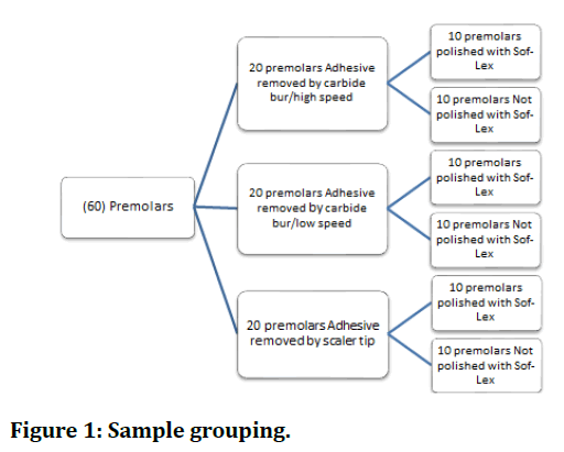
The buccal enamel surfaces of 60 premolar teeth were bonded with metal brackets then were divided randomly into three into 3 categories, each with twenty teeth according to the technique of adhesive removal. Every group included ten teeth that were polished with Sof-lex polishing discs and ten teeth that were not polished with Sof-lex polishing discs. This results in six subgroups, the high speed carbide bur followed by Sof-lex polishing disks (HSP), the high speed carbide bur without polishing (HS), low speed handpiece carbide bur followed by Sof-lex polishing disks (LSP), low speed handpiece carbide bur without polishing (LS), sickle scalar’s tip in ultrasonic scalar followed by Sof-lex polishing disks (USP) and sickle scalar’s tip in ultrasonic scalar without polishing (US), as shown in the Figure 1.

Figure 1. Sample grouping.
The teeth used were extracted for general dental reasons and were collected from private dental clinics. Instantly after extraction, teeth were debrided by water to remove soft tissue remnants, debris, or blood, and examined under a stereomicroscope (LeicaTM, Leitz, Wetzlar, Germany) at a tenfold magnification to verify that they were generally healthy and free from caries, restorations, enamel cracks, or surface imperfections were found, and there was no previous endodontic, orthodontic, or bleaching treatment history. Then, for a maximum of one week, store in a 1% chloramine-T trihydrate bacteriostatic/bactericidal solution, followed by storage in distilled water to avoid dehydration [11].
Sample Preparation
Each tooth will be mounted vertically on a glass slide with soft sticky wax at the end of the root, so that the middle third of the buccal surface is parallel to the surveyor's analyzing rod. The custom-made cylindrical mold will next be painted with a thin layer of separating medium (artificial saliva) and put around the teeth in a vertical posture with crowns protruding. The cold cured acrylic powder and liquid will then be mixed and applied around the teeth to the cemento-enamel junction of each tooth.
After mounting, the specimens were coded from (1-60) each sample had its specific number at the base of the acrylic and stored in deionized water in closed containers. The deionized water was replaced every day until the day of bonding to prevent dehydration and bacterial growth.
Each tooth had a rectangular piece of adhesive tape placed over the middle third of the buccal surface. An opening (round in shape) was left on the tape for standardization, allowing bonding, adhesive removal, and color analysis with the spectrophotometer [12].
A mold was made from plaster similar in shape to the base of orthodontic study model. The mold had central hole to allow the acrylic block to be placed and fixed in the dental surveyor table during polishing, adhesive removal and color measurement of each tooth and then removed and replaced with another tooth. Each tooth's buccal surface was polished with no fluoridated pumice and a rubber cup. For this study's standardization, one rubber cup was placed to a slow speed hand piece for 10 seconds polishing for each tooth. After that, each tooth was sprayed with water for 10 seconds before being dried with oil-free air for 10 seconds. The air water syringe was fixed at 1 cm away from the buccal tooth surface for standardization.
Bonding Procedure
At room temperature, the priming procedure was performed as the following:
The traditional acid etching approach was used to bond all of the premolars. The enamel was etched for 30 seconds with 37 percent phosphoric acid, and then rinsed with water spray for 10 seconds before being dried with oil-free air for 20 seconds. A thin film of Transbond XT primer was painted on the etched enamel surface with gingivaocclusal direction using a disposable brush, then polymerized by a LED light curing unit (Curing intensity 1300 mw/cm2) for 10 seconds.
Each sample received a maxillary first premolar bracket, with the mesh bonding surface coated with separating media (artificial saliva) before being coated with composite resin adhesive. The bracket was then positioned in the middle third of the buccal surface, parallel to the long axis of the teeth, and pushed toward the tooth surface with clamping tweezers.
To guarantee that each bracket was seated under an equal pressure and to establish a uniform thickness of the adhesive, a steady load (200 gm) was applied on the bracket for 10 seconds immediately after bracket positioning, and the extra material was removed using a sickle probe.
In all groups, the bracket adhesive was exposed to the curing light for 40 seconds (20 seconds from the mesial and 20 seconds from the distal according to the manufacturer's instructions) at a distance of 5 mm using a ruler fixed at the tip of the light cure device for standardization and angulated at 45 degrees to the proximal sides of the bracket. The intensity of curing light was 1300 mw/cm2 and it was rechecked periodically via curing light meter before usage.
The bonded teeth of all groups were stored in an incubator in distilled water inside sealed containers at 37°C for 24hr [11].
Each bracket was then removed by using a clamping tweezers creating a relatively standardized bonded composite resin rectangle that resembles the shape of maxillary first premolar bracket base.
Removal of adhesive
The adhesive was cleaned by using the procedure specified for each group. For each sample in groups HSP, HS, LSP, and LS, a new bur was used, however in groups USP and US, the same scalar tip was used for all samples. The removal of the composite was considered done when seen under the illumination of the operatory lamp and the tooth surface seemed free of composite to the naked eye, equivalent to the clinical work [13,14].
After that, medium, fine, and ultra-fine Sof–Lex XT discs (3M ESPE, St Paul, Minnesota) were used to polish the teeth in groups (HSP), (LSP), and (USP). New discs were used for each sample [9]. Polishing time and sequence were standardized for each tooth that was polished; each disc was used for 10 seconds [15].
Staining procedure
One teabag was added to 250 mL of freshly boiled water and stirred for 5 minutes before removing the bag. A quantity of 250 ml was chosen since it was the average volume of a regular tea mug [16]. The tea was allowed to cool until it approached 55°C prior to teeth immersion.
All samples were immersed in the prepared tea daily for 10 minutes then washed and stored in distilled water at 37ºC for the reminder of the day. The process continued for 30 days [16].
Spectrophotometer measurement
The VITA easyshade® spectrophotometer was used to take specttrophotometric color measures for all the teeth initially (before bonding), these were recorded as first measurement (E1). The bond removal procedure specific for each group was conducted as previously mentioned, followed by a second measurement for all samples (E2). Then a third measurement after 30 days of immersion of all teeth in tea will be recorded as (E3). Prior to measuring a tooth color the VITA easy shade was calibrated according to the manufacturer's instructions (Place the device in the calibration block holder so that the probe tip is flush with and perpendicular to the calibration block, and the calibration is depressed, and the handpiece is fully inserted in the calibration holder). The calibration was repeated before every measurement.
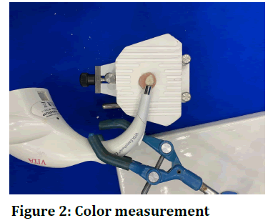
The probe of the Spectrophotometer was placed at 90 ° contacting the buccal surface of the tooth in the intended bracket position to record the tooth color (Figure 2). To reduce the margin of error, three measurements were taken, and the average of the three measurements was determined. The CIE (Commission Internationale de l'Eclairage) L*a*b* color system (1931) was used to evaluate color, which uses three factors to determine color: The L* coordinate represents the degree of lightness and darkness and runs from 0 to 100 (white), while the a* and b* coordinates represent places on the red (+)/ green (-) and yellow (+)/ blue (-) axes, respectively. The variation between two colours was determined (the distance between the 2 points in color space) with the following formula:

Figure 2. Color measurement
ΔE={(L2-L1)2 (a2-al)2 (b2-b1)2}1/2 [9].
ΔE 1: The variation between the values obtained at the starting of orthodontic therapy before bonding and after adhesive removal (baseline-adhesive removal). Clinically, this number represents the discoloration throughout orthodontic therapy.
ΔE 2: The variation between the values obtained after cleaning the surfaces and the final values after immersion in tea for 30 days (adhesive removal-final), and clinically represents the discoloration that occurs after the therapy.
ΔE of each group was compared with those of the others, to distinguish which type of adhesive removal procedures was more unstable in color, and to evaluate the effectiveness of Sof-Lex polishing discs on tooth color stability.
Statistical analysis
Statistical analysis of data was performed using SPSS StatisticsTM software version 26.0 (IBM Company, New York, USA). Normality of data distribution was tested using the Shapiro-Wilk test, which showed that ΔE values were normally distributed. Analysis of statistical differences was carried out using ANOVA for ΔE values. The significance level was set at 𝑃≤ 0.05.
Results
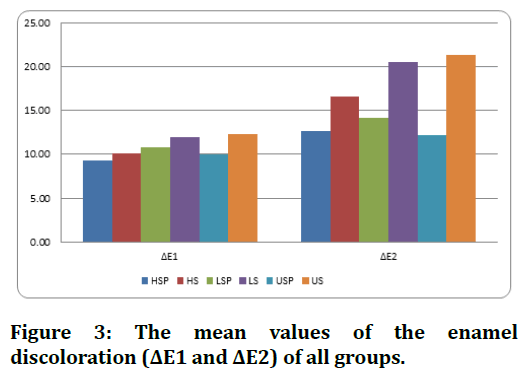
The means of ΔE values for each group are given in (Table 1) and (Figure 3).
| Discoloration | Group | No | Mean |
|---|---|---|---|
| ΔE1 | HSP | 10 | 9.25 |
| HS | 10 | 10.08 | |
| LSP | 10 | 10.8 | |
| LS | 10 | 11.94 | |
| USP | 10 | 10.04 | |
| US | 10 | 12.33 | |
| ΔE2 | HSP | 10 | 12.66 |
| HS | 10 | 16.65 | |
| LSP | 10 | 14.2 | |
| LS | 10 | 20.55 | |
| USP | 10 | 12.24 | |
| US | 10 | 21.36 |
Table 1: Descriptive statistics of the ΔE values of different groups

Figure 3. The mean values of the enamel discoloration (ΔE1 and ΔE2) of all groups.
Discoloration from baseline to adhesive removal (ΔE1)
The highest mean value of ΔE1 was in US group (12.33 ± 3.17), followed by that of LS group (11.94 ± 2.89), then LSP group (10.80 ± 3.78), then HS (10.08 ± 1.87), then USP group (10.04 ± 1.63) and lastly the HSP group, which had the lowest mean of ΔE1 (9.25 ± 1.00).
Discoloration from adhesive removal to final record (ΔE2)
The highest mean value of ΔE2 was in US group (21.36 ± 6.53), followed by that of LS group (20.55 ± 2.17), then HS group (16.65 ± 3.45), then LSP (14.20 ± 3.14), then HSP group (12.66± 3.93) and lastly the USP group, which had the lowest mean of ΔE2 (12.24± 2.73).
The comparison of mean differences in enamel color changes among all groups at ΔE1 and ΔE2 were determined using One-way analysis of variance (ANOVA) test and showed both significant and non-significant differences. The Post-hoc Tukey’s test was carried out for multiple comparisons to reveal the differences among the tested groups (Table: 2).
| Discoloration | ANOVA test | ||
|---|---|---|---|
| F-test | P-value | ||
| ΔE1 | Between Groups | 1.756 | 0.138 (NS) |
| ΔE2 | Between Groups | 10.16 | 0.000 (VHS) |
Table 2: ANOVA test for the comparison among the groups regarding (ΔE 1, ΔE 2)
The results showed statistically no significant difference among the adhesive removal techniques when compared according to ΔE1, but there was a very highly significant difference among these techniques when compared according to ΔE2 (p value was 0.000).
In order to verify between which adhesive removal technique was the difference; Post Hock Tukey’s test was performed (Table 3). The results shows a statistically very highly significant difference between HSP and LS groups, HSP and US groups, LS and USP groups, and USP and US groups (p value was 0.000), while there were significant differences between LSP and LS groups and LSP and US and a non-significant difference between HSP and HS groups, HSP and LSP groups, HSP and USP groups, HS and LSP groups, HS and LS groups, HS and USP groups, HS and US groups, LSP and USP groups and LS and US groups.
| Dependent Variable | Between Subgroups | P value | |
|---|---|---|---|
| ΔE 2 | HSP | HS | .221 (NS) |
| LSP | .950 (NS) | ||
| LS | .000 (VHS) | ||
| USP | 1.000 (NS) | ||
| US | .000 (VHS) | ||
| HS | LSP | .727 (NS) | |
| LS | .244 (NS) | ||
| USP | .137 (NS) | ||
| US | .094 (NS) | ||
| LSP | LS | .008 (HS) | |
| USP | .871 (NS) | ||
| US | .002 (HS) | ||
| LS | USP | .000 (VHS) | |
| US | .997 (NS) | ||
| USP | US | .000 (VHS) | |
Table 3: Comparison of color change between the values obtained after cleaning the enamel surfaces and the final values after immersion of teeth in tea.
Discussion
The correlation between tooth discoloration and orthodontic treatment using fixed appliances remains debatable. According to some researchers who concluded that bonding and debonding methods alone did not seem to have a significant effect on the color of human tooth enamel. On the other hand, several researchers have found that these methods have a considerable impact on enamel color parameters [5]. In daily practice, the change in enamel color after orthodontic treatment is frequently neglected. The color of tooth enamel varies in a variety of ways after thorough orthodontic treatment with fixed appliances [17]. After debonding the orthodontic appliances, the CIE color parameters L*, a*, and b* of natural teeth exhibited statistically significant alterations. Permanent iatrogenic enamel effects involved with bonding, debonding, and cleaning treatments; exogenous and endogenous discoloration of the residual adhesive material; and dental and pulp tissue modifications linked with orthodontic tooth movement could all contribute to this result. External colouring happens as a consequence of superficial absorption of dietary dyes, whereas internal colouring develops as a result of aging [2]. The limitations of this methodology include the absence of saliva, the food colouring, and the inability to mimic the mechanical abrasion induced by brushing
Fixed orthodontic treatment, without a doubt, produces irreparable damage to teeth enamel. When removing adhesives, two types of iatrogenic enamel damage should be regarded: enamel loss caused by etching, grinding, and subsequent polishing, and enamel roughening caused by scratching or faceting [18].
The final enamel appearance following debonding should, for all practical purposes, be comparable to the neighboring natural enamel surfaces, both dry and wet. Because reflection and refraction processes associated with a wet surface might disguise surface defects, it's crucial to examine the dry appearance [19].
To restore the enamel surface, it was advised that once remaining adhesive resins were removed, the enamel surface should polished [20,21]. This was consistent with [19] who observed that Sof-Lex finishing and polishing disks with different grades of medium, fine and superfine provided surfaces that could be easily restored after the final polish.
Tungsten carbide burs are faster and more effective at removing adhesive than Sof-Lex discs, ultrasonic tools, hand instruments, rubbers, or composite burs. They remove a significant layer of enamel and roughen its surface, therefore multi-step Sof-Lex discs and pumice slurry, which is the most efficient way of polishing, should be used afterward [18].
Following clean-up techniques, the presence of remnant adhesive resin in the enamel surface of the middle third of the tooth induced color instability, which was induced by direct absorption of external colorants [21].
The use of a high-speed tungsten carbide bur or an ultrasonic scalar produced the most enamel loss. The use of a slow-speed tungsten carbide bur or debonding pliers resulted in the least amount of enamel loss [22], while another study suggested that a high-speed tungsten carbide bur, multiple grades of Sof-Lex discs, and lastly zircate paste must be used [19].
The quality of post debonding clean-up techniques, which could remove remaining adhesive and restore tooth aesthetic appearance, had a big impact on enamel discoloration. However, grinding with various rotary devices that exacerbated tooth color change, particularly rough clean-up procedures and/or instruments, such as employing a diamond bur, resulting in unavoidable enamel surface loss and abnormalities [23].
In the present study, enamel color changes after debonding and cleaning procedures in all groups, the color difference was more than the perceptible limit (ΔE>3.7 units) and ranged from 9.25 to 12.33 ΔE units. These findings corroborate to several studies that study the impact of orthodontic bonding on the color of the teeth [1,2,8,9,17].
The large differences observed for baseline-adhesive removal could be explained by the extensive nature of the debonding and adhesive cleaning related techniques. During acid etching, the apatite crystallites dissolve, increasing the microscopic roughness of the exposed enamel [24] and, as a result, the light scattering of the tooth surface [25]. Furthermore, Present bracket debonding and adhesive removal techniques frequently cause enamel morphology and surface changes that cannot be restored by polishing during the post finishing steps [17].
There were no statistically significant group differences among the color changes baseline-adhesive removal mechanisms. The absence of statistically significant differences with respect to ΔE1 among the adhesive removal mechanisms implies that the influence of the surface roughness produced by cleaning and polishing techniques may outweigh any variations in the composition of enamel surfaces subjected to debonding. This observation is important for orthodontists who may adversely influence the roughness of the bonding surface by grinding the enamel during adhesive removal [8].
In the present study, enamel color changes that occurred between adhesive removal to the final measurement after immersion in tea procedures in all groups, the color difference was more than the perceptible limit (ΔE>3.7 units) and ranged from 12.36 to 21.36 ΔE units.
When comparing the results of enamel discoloration among all adhesive removal techniques used in this study, less enamel discoloration observed in the groups that include polishing than that without polishing groups. This finding could be explained by the capability of the graded abrasive discs to eliminate the surface scratches produced by the adhesive removal carbide burs or scalar’s tips and restore the tooth surface [23]. It was reported that the polishing of enamel following adhesive removal show less stain susceptibility [21, 23].
Conclusion
All techniques of adhesive removal showed that there was visible and clinically significant alterations in color, larger than the value of clinical detection threshold (ΔE>3.7 units). Less enamel discoloration observed in the groups that include polishing than that without polishing groups. USP and HSP groups are the least stain susceptibility while the US group show the highest discoloration after immersion in tea.
References
- Al Maaitah EF, Omar AA, Al-Khateeb SN. Effect of fixed orthodontic appliances bonded with different etching techniques on tooth color: a prospective clinical study. Am J Orthod Dentofacial Orthop 2013; 144:43-9.
- Boncuk Y, Cehreli ZC, Polat-Özsoy Ö. Effects of different orthodontic adhesives and resin removal techniques on enamel color alteration. Angle Orthod 2014; 84:634-41.
- Sundfeld RH, Franco LM, Machado LS, et al. Treatment of enamel surfaces after bracket debonding: case reports and long-term follow-ups. Oper Dent 2016; 41:8-14.
- Leland A, Akyalcin S, English JD, et al. Evaluation of staining and color changes of a resin infiltration system. Angle Orthod 2016; 86:900-4.
- Chen Q, Zheng X, Chen W, et al. Influence of orthodontic treatment with fixed appliances on enamel color: A systematic review. BMC Oral Health 2015; 15:1-8.
- Judeh A, Al-Wahadni A. A comparison between conventional visual and spectrophotometric methods for shade selection. Quintessence Int 2009; 40:e69-79.
- Brewer JD, Wee A, Seghi R. Advances in color matching. Dent Clin 2004; 48:341-358.
- Eliades T, Kakaboura A, Eliades G, et al. Comparison of enamel colour changes associated with orthodontic bonding using two different adhesives. Eur J Orthod 200 ;23:85-90.
- Gorucu–Coskuner H, Atik E, Taner T. Tooth color change due to different etching and debonding procedures. Angle Orthod 2018; 88:779-84.
- George J, Mathews TA. Spectrophotometric Analysis of tooth discoloration after bracket debonding induced by coffee and tea grown in South India: An In vitro Study. J Interdiscip Dent 2018; 8:1.
- Ibrahim AI, Thompson VP, Deb S. A novel etchant system for orthodontic bracket bonding. Scientific Reports 2019; 9:1-5.
- Ijaz U, Haroon K, Haroon S, et al. Enamel surface roughness assessment after debonding, employing three different removal methods. Profe Med J 2019; 26:1511-7.
- Zaher AR, Abdalla EM, Abdel Motie MA, et al. Enamel colour changes after debonding using various bonding systems. J Orthod 2012; 39:82-8.
- Janiszewska-Olszowska J, Tomkowski R, Tandecka K, et al. Effect of orthodontic debonding and residual adhesive removal on 3D enamel microroughness. Peer J 2016; 4:e2558.
- Koh R, Neiva G, Dennison J, et al. Finishing systems on the final surface roughness of composites. J Contemp Dent Pract 2008; 9:138-45.
- Nahidh M. The effects of various beverages on the shear bond strength of light-cured orthodontic composite (An in vitro comparative study). J Baghdad Coll Dent 2014; 26:144-148.
- Karamouzos A, Athanasiou AE, Papadopoulos MA, et al. Tooth-color assessment after orthodontic treatment: a prospective clinical trial. Am J Orthod Dentofacial Orthop 2010; 138:537-e1.
- Janiszewska-Olszowska J, Szatkiewicz T, Tomkowski R, et al. Effect of orthodontic debonding and adhesive removal on the enamel–current knowledge and future perspectives–a systematic review. Med Sci Monit: Int Med J Exp Clin Res 2014; 20:1991.
- Zarrinnia K, Eid NM, Kehoe MJ. The effect of different debonding techniques on the enamel surface: An in vitro qualitative study. Am J Orthod Dentofacial Orthop 1995; 108:284-93.
- Zachrisson BU, Årthun J. Enamel surface appearance after various debonding techniques. Am J Orthod 1979; 75:121-37.
- Joo HJ, Lee YK, Lee DY, et al. Influence of orthodontic adhesives and clean-up procedures on the stain susceptibility of enamel after debonding. Angle Orthod 201; 81:334-40.
- Hosein I, Sherriff M, Ireland AJ. Enamel loss during bonding, debonding, and cleanup with use of a self-etching primer. American J Orthod Dentofacial Orthop 2004; 126:717-24.
- Ye C, Zhao Z, Zhao, Q et al. Comparison of enamel discoloration associated with bonding with three different orthodontic adhesives and cleaning-up with four different procedures. J Dent 2013; 41:e35-40.
- Retief DH, Busscher HJ, De Boer P,et al. A laboratory evaluation of three etching solutions. Dent Mat 1986; 2:202-6.
- Ten Bosch JJ, Coops JC. Tooth color and reflectance as related to light scattering and enamel hardness. J Dent Res 1995; 74:374-80.
Author Info
Hasan Muneer Raoof* and Nidhal Hussien Ghaib
Department of Orthodontics, College of Dentistry, University of Baghdad, IraqCitation: Hasan Muneer Raoof, Nidhal Hussien Ghaib,Evaluation of Enamel Discoloration Following Different Orthodontic Adhesive Removal Procedure: An in Vitro Study, J Res Med Dent Sci, 2021, 9(12): 117-123
Received: 04-Oct-2021 Accepted: 24-Nov-2021
