Case Series - (2021) Volume 9, Issue 7
Oncoplastic Surgical Technique in Granulomatous Mastitis-A Case Series
Manish Kaushal and Aditi Sharma*
*Correspondence: Aditi Sharma, Department of General Surgery, Mahatma Gandhi Memorial Medical College and Maharaja Yashwantrao Hospital, India, Email:
Abstract
Introduction: Granulomatous mastitis is a chronic inflammatory lesion of the breast with varying presentation. It is a benign etiology with high recurrence rates that mimic breast malignancy. Well defined guidelines for the treatment of granulomatous mastitis do not exist but most recommended treatment includes medical management with antibiotics and steroids and wide surgical excision of the diseased tissue. We here discuss excision of the breast lump using Oncoplastic techniques thus providing good aesthetics. Methods: 6 patients who presented to our OPD with breast lumps and fistula were made a diagnosis of GM by exclusion. Wide excision with the application of therapeutic mammoplasty principles were used in each of them and patients were followed for 6 months post op to see the results. Outcome: All 6 patients showed remission with no recurrence 6 months post op and a good breast appearance. Conclusion: Oncoplastic surgical techniques in the excision of granulomatous mastitis is recommended with good aesthetic outcome.
Keywords
Granulomatous mastitis, Oncoplastic mammoplasty, Donut mastopexy, Case series
Introduction
Granulomatous mastitis is a chronic inflammatory lesion of the breast characterized by granulomatous changes around the ducts and lobules of breast either of unknown etiology or secondary to corny bacterium, tuberculosis, fungal infection or connective tissue disorders like Wegener’s disease and sarcoidosis. When of unknown etiology, the term idiopathic granulomatous mastitis is used which was first described by Kessler and Wolloch in 1972 [1]. IGM can mimic inflammatory breast cancer, clinically as well as radio logically. Diagnosis is mainly one of exclusion and histopathology forms the basis for the diagnosis of IGM [2].
The optimal treatment guidelines for granulomatous mastitis are still a matter of discussion. Usually, these patients present with painful breast lumps and abscess with or without skin ulceration/ fistula. There is history of recurrent abscess drainage with multiple scars and long intake of oral antibiotics without much benefit. Surgical management consists of wide excision of diseased breast with the best cosmetic outcome. Oncoplastic breast surgical techniques and reconstruction have shown a good result in these patients. Here we discuss 6 patients and surgical excision of the breast lump using Oncoplastic surgical techniques.
Materials and Methods
This is a prospective case series conducted over a period of 2 years in a single centre. Six patients in the age group 26-46 years presented to our OPD with unilateral recurrent mastitis and multiple abscess formation. Baseline characteristics such as age at diagnosis, history of pregnancy and lactation, history of TB or autoimmune disease, oral contraceptive use, smoking status, medical ailment and family history of breast cancer were noted for all the patients.
Primary symptoms, the presence of a mass, abscess, or fistula, location of the lesion within the breast, and nipple involvement were evaluated. Diagnostic studies including culture studies, mammograms, ultrasounds, and histologic examinations were performed. Patients with large breast lumps and with involvement of nipple areola complex were not included in this study.
A provisional diagnosis of granulomatous mastitis was made in all 6 cases by exclusion. These patients had been taking antibiotics since a few weeks and had undergone multiple drainage procedures with failure to treat the disease. 2 of the 6 patients had received anti tubercular treatment for 6 months with no clinical improvement. Surgical techniques like wide local excision, partial mastectomy with lateral extension and donut mastopexy were performed in these patients under antibiotic coverage. Post operatively, the patients were followed for 6 months to look for any recurrences and failure of treatment.
Results
All the 6 patients were in the reproductive age group with none being pregnant or lactating at the time of presentation. None of the patients had history of smoking and oral contraceptive pills. No history of medical disease or tuberculosis was noted in any patient.
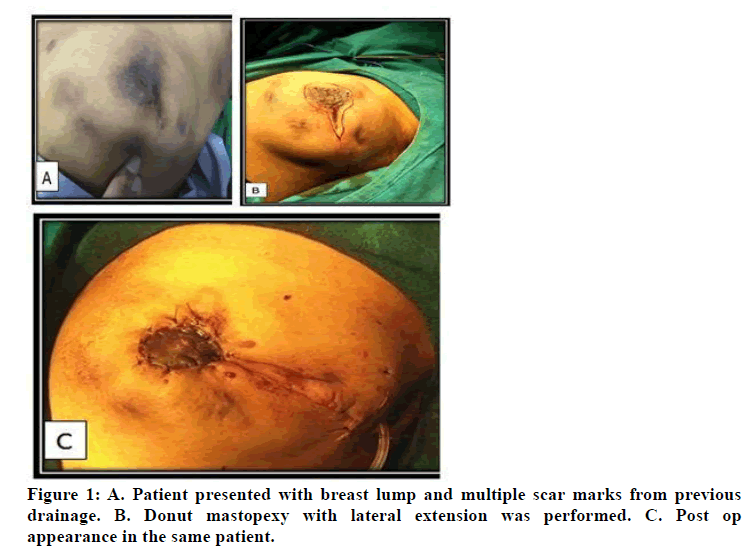
Four patients presented with unilateral breast lump with scar marks of previous drainage. 1 patient had presented with fistulous scar and discharge. 1 patient had breast pain with indurated region.
Ultrasound reported presence of hypo echoic irregular lesions of BIRADS IV category. Core needle biopsy confirmed granulomatous mastitis in 2 patients and 4 reports were inconclusive. Post op histopathological reports showed IGM in all cases with one showing tuberculous etiology. Pus culture and fungal cultures failed to how any organism in any case.
The procedures performed were lateral mammoplasty with partial mastectomy, donut mastopexy with lateral extension and wide local excision.
Patients were subjected to surgical excision of the mass according to the location of the mass within the breast. Figure 1 shows the pre-operative, surgical incision and immediately post op image of a patient. Patients showed uneventful recovery without any complications. No recurrence was seen after mastopexy and wide local excision till 6 months post op. Disease remission with good cosmesis was obtained for all these patients.

Figure 1: A. Patient presented with breast lump and multiple scar marks from previous drainage. B. Donut mastopexy with lateral extension was performed. C. Post op appearance in the same patient.
Discussion
IGM is a rare chronic inflammatory disease of the breast which affects women of child-bearing age. It is more common in the developing world and is often associated with pregnancy and lactation [3,4]. The prevalence and incidence of the disease have been reported as 2.4 per 100,000 women and 0.37%, respectively [5,6].
IGM can present as a rapidly increasing breast mass, which may be associated with inflammation, nipple retraction, abscesses and chronic draining sinuses [7]. There is a tendency for the condition to recur, and patients with recurrent sterile breast abscesses should have IGM excluded [8].
Diagnosis is mainly one of exclusion and can only confirm by histopathology [9]. It shows the features of a noncaseating granuloma, composed of epithelioid histiocytes with giant cells within and around the lobules [10].
The natural history of GLM may be self-limited, and expectant management with close surveillance has been suggested but since most cases are accompanied by complications such as an abscess or fistula, using expectant management or antibiotics alone is not effective. Steroids have a proven benefit in the management of these patients since autoimmune aetiology is suspected in idiopathic cases. The use of corticosteroids for the treatment of idiopathic granulomatous mastitis (IGM) was first proposed by DeHertogh et al. [11]. Many studies thereafter have been conducted proving the role of steroids in remission of IGM. The use of steroids reduces the dimensions of the disease and can help to promote healing after excision of the disease [12,13]. The exact duration and dose of steroid is still a matter of debate.
In our study, we did not use steroids as initial treatment and was reserved for recurrent cases. Surgical management initially comprised of incision and drainage of abscess and local excision of diseased tissue. This was associated with high recurrence rates thus wide local excision including the fistulous tract is now practised. Although recurrences are known to occur even after wide excision, it has become treatment of choice. According to Hur et al. surgical excision showed a fast recovery rate, and high success rate (90.3%) with low recurrence (8.7%) [14]. This is supported by Atak et al. [15].
In the recent years, a multidisciplinary team approach is followed which consists of both breast and plastic surgeons to provide both adequate excision as well as good aesthetic reconstruction of the breast. The use of Oncoplastic breast reconstructive techniques has been utilized and supported by various studies. It plays a fundamental role in the management of patients afflicted with IGM, as it gives the surgeon the freedom to perform a wider resection, thus reducing the rate of recurrence, whilst at the same time, knowing that there are reconstructive options to restore any breast aesthetics [16]. Wide excision of breast tissue or mastectomy followed by immediate or delayed breast reconstruction is offered to the patients. Using therapeutic mammoplasty techniques in surgical management of IGM in moderate to large breasts seems justifiable with good results regarding recurrence and postoperative patients’ satisfaction [17].
In our study, we followed the Oncoplastic principles of breast tissue excision. Surgeries such as donut mastopexy with lateral and medial extension were performed in 6 patients. None of these showed any recurrence up to 6 months post op. The patients were satisfied with the appearance of post op breast.
Conclusion
The optimal management of GM is still controversial. The role of I & D is limited because it may not improve the condition and is associated with a high rate of recurrence with poor cosmetic result. It may lead to intractable incision tracks, which subsequently leads to sinus formation.
Using therapeutic mammoplasty techniques in surgical management of GM in moderate to large breasts provides good results in terms of recurrence and cosmesis, while providing adequate amount of tissue for histopathological assessment for reaching a confirmatory diagnosis of GM.
References
- Kessler E,Wolloch Y. Granulomatous mastitis: a lesion clinically simulating carcinoma. Am J Clin Pathol 1972; 58:642–646.
- Erhan Y, Veral A, Kara E, et al. A clinicopthologic study of a rare clinical entity mimicking breast carcinoma: idiopathic granulomatous mastitis. Breast 2000; 9:52–56.
- Bani-Hani KE, Yaghan RJ, Matalka II, et al. Idiopathic granulomatous mastitis: time to avoid unnecessary mastectomies. Breast J 2004; 10:318–322.
- Going JJ, Anderson TJ, Wilkinson S, et al. Granulomatous lobular mastitis. J Clin Pathol 1987; 40:535–540.
- Centers for Disease and Prevention. Idiopathic granulomatous mastitis in Hispanic women - Indiana, 2006-2008. Morb Mortal Wkly Rep 2009; 58:1317–1321.
- Ahmed R, Sultan F. Granulomatous mastitis: a review of 14 cases. J Ayub Med Coll Abbottabad 2006; 18:52–54.
- Yabanoğlu H, Çolakoğlu T, Belli S, et al. A comparative study of conservative versus surgical treatment protocols for 77 patients with idiopathic granulomatous mastitis. Breast J 2015; 21:363-9.
- Sheybani F, SarvghadMR, Naderi HR, et al. Treatment for and clinical characteristics of granulomatous mastitis. Obstet Gynecol 2015; 125:801–807.
- Oran ES, Gürdal SÖ, Yankol Y, et al. Management of idiopathic granulomatous mastitis diagnosed by core biopsy: A retrospective multicenter study. Breast J 2015; 19:411–418.
- Gurleyik G, Aktekin A, Aker F, et al. Medical and surgical treatment of idiopathic granulomatous lobular mastitis: A benign inflammatory disease mimicking invasive carcinoma. J Breast Cancer 2012; 15:119–123.
- DeHertogh DA, Rossof AH, Harris AA, et al. Prednisone management of granulomatous mastitis. N Engl J Med 1980; 303:799–800.
- Asoglu O, Ozmen V, Karanlik H, et al. Feasibility of surgical management in patients with granulomatous mastitis. Breast J 2005; 11:108–114.
- Akcan A, Akyıldız H, Deneme MA, et al. Granulomatous lobular mastitis: A complex diagnostic and therapeutic problem. World J Surg 2006; 30:1403–1409.
- Hur SM, Cho DH, Lee SK, et al. Experience of treatment of patients with granulomatous lobular mastitis. J Korean Surg Society 2013; 85:1–6.
- Atak T, Sagiroglu J, Eren T, et al. Strategies to treat idiopathic granulomatous mastitis: retrospective analysis of 40 patients. Breast Dis 2015; 35:19–24.
- McLean NR, Chummun S, Youssef MK, et al. Delayed breast reconstruction in idiopathic granulomatous mastitis. Eur J Plast Surg 2019; 42:243–249.
- Ahmed YS, Abd El Maksoud W. Evaluation of therapeutic mammoplasty techniques in the surgical management of female patients with idiopathic granulomatous mastitis with mild to moderate inflammatory symptoms in terms of recurrence and patients satisfaction. Breast Disease 2016; 36:37-45.
Author Info
Manish Kaushal and Aditi Sharma*
Department of General Surgery, Mahatma Gandhi Memorial Medical College and Maharaja Yashwantrao Hospital, Indore, Madhya Pradesh, IndiaCitation: Manish Kaushal, Aditi Sharma,Oncoplastic Surgical Technique in Granulomatous Mastitis-A Case Series, J Res Med Dent Sci, 2021, 9(7): 225-227
Received: 24-Mar-2021 Accepted: 13-Jul-2021
