Review Article - (2023) Volume 11, Issue 8
Salivary pH as a Diagnostic Marker in Oral Sub-Mucous Fibrosis
Nelakurthi Vidya Maheswari* and Aarati Panchbhai
*Correspondence: Dr. Nelakurthi Vidya Maheswari, Department of Oral Medicine and Radiology, Datta Meghe Institute of Medical Sciences, Maharashtra, India, Email:
Abstract
Oral sub mucous fibrosis precancerous, long standing, progressive disease of oral cavity containing fibrous bands in oral mucosa. Various factors have been predicted to cause the changes in salivary pH. Habitual areca nut and tobacco chewing are one of the most important causes of salivary pH changes in oral sub mucous fibrosis. As saliva is easily collected from oral cavity it can be used as diagnostic marker for many diseases. Saliva contains an enzyme superoxide dismutase that has antioxidative property. Long term intake of tobacco and its substances decrease the activity of this enzyme. Present review concludes that there is no account alteration in salivary pH of normal individuals and oral sub mucous individuals however, the salivary pH of oral sub mucous fibrosis patients is slightly alkaline as compared to normal healthy individuals after habitual chewing substance.
Keywords
Oral submucous fibrosis, Salivary pH, Baseline salivary pH, Post chew salivary
Introduction
Oral submucous fibrosis a longlasting juxta-epithelial inflammatory disease of oral cavity. It is the precancerous condition with progressive scarring which contains fibrous bands in the oral mucosa. Owing to pre malignant condition of oral cavity it has high incidence of converting to malignancy [1,2]. Oral submucous fibrosis has now days accepted and regarded as Indian disease as the prevalence increased from 0.03% to 6.42%. Due to the crippling nature of the disorders, the studies are focused on exploring its nature in terms of etiology, risk factors and management. In oral sub mucous fibrosis, the patient present with clinical features of difficulty in opening mouth, stiffness of labial and buccal mucosa, burning sensation of mucosa on taking spicy food is the earliest symptom. In severe cases, there is difficulty in swallowing, speech and xerostomia can be observed. It is mainly caused due to irritation of oral mucosa due to areca nut which is considered as the most predominating contributing factor. Tobacco, gutkha, kharra, pan masala, khaini, snuff etc. cause the release of dangerous chemicals that causes changes in salivary pH. Areca nut when taken along with lime which contains calcium oxide and calcium hydroxide or tobacco chewing provides more alkaline pH. Alkaline exposure in the oral cavity is compensated by fibrous bands formation [3].
Areca nut contains four main alkaloids such as arecoline, arecadine, guvacoline and guvacine which have vasoconstricting properties. The constituents of tobacco include nicotine, CO, tar, nitrogen oxides, HCN. Histopathological features of oral sub mucous fibrosis include excessive collagen bundle deposition in the lamina propria and chronic inflammatory cell infiltrate is present below the oral epithelium and atrophy of the oral epithelium because of this reason scoring of epithelial dysplasia of oral cavity is not feasible. Clinically oral sub mucous fibrosis shows white bands which can be palpated by fingers. Clinically, fibrosis can be mostly felt on buccal mucosa, labial mucosa and soft palate. The use of areca nut increases the prevalence of premalignant condition, malignancy and other diseases and it was also playing a major role in public health issues [4].
Literature Review
Siddabasappa, et al has done a study to assess and compare salivary flow rate, salivary pH, iron, copper of gutkha chewers who are free or suffering from oral submucous fibrosis. The study included 60 individuals among which 20 are ghutka chewers having oral sub mucous fibrosis, 20 is ghutka chewers without oral sub mucous fibrosis and 20 are gender matched normal individual, salivary pH was assessed by Elico-L1-612 pH analyser. The result stated that salivary pH was not altered. Tamgadge and Wasekar has conducted a case control study to evaluate and compare salivary pH. The study was conducted on 90 individuals among which they were divided into three groups, 30 patients are gutka chewers with oral sub mucous fibrosis, 30 patients are gutka chewers without oral submucous fibrosis and 30 individuals are controls who are healthy individuals. The results shows that the changes in salivary pH in three groups is not significant [5].
Sushmitha S et al., has conducted a study to know the effects of tobacco and tobacco products on salivary pH, taste perception and salivary flow rate in OSMF and leukoplakia patients. The study inclulded 45 patients with an age group of 20-50 years. The individuals are divided into three groups as OSMF patients, leucoplakia and normal individuals. The salivary flow rate, salivary pH and taste perception is assessed by using schirmer strips, pH strips and taste strips respectively. The result states the there is a definite change in these parameters in OSMF and leucoplakia patients when compared with normal individuals. OSMF patients show greater alterations when compared with leucoplakia patients [6].
A study investigated salivary flow rate, salivary pH and taste perception in 90 individuals in which 45 are OSMF patients and 45 are normal individuals. The three parameters were compared among stage I (14) and stage II (31) OSMF patients. Study concluded that no accountable change in the mean salivary pH between OSMF patients and normal individuals and the salivary flow rate is decreased in oral submucous fibrosis patients than normal individuals and taste perception to salty and sour in OSMF stage I when compared to stage II. A study was done among habitual areca nut and various tobacco product consumers to assess the salivary pH changes, the 360 individuals (chewers and non-chewers) have been enrolled in the study with an age group between 20-30 years. Baseline and post chew salivary pH of the patient were collected and measured by using digital pH meter. Results in all chewers groups were of decrease in pH except in gutkha and lime chewing group where the pH is increased [7]. The mean pH of chewers has been increased as compared to the mean pH of normal individuals. Shubha et al., estimated salivary flow rate and salivary pH in smoking and smokeless form of tobacco in comparison of salivary pH. A statistically significant decrease is seen in smokeless tobacco group than with control group. A accountable reduction in salivary flow rate and slight decrease in salivary pH was detected. Faisal Reehan, in his study assessed resting salivary flow rate and pH in 210 tobacco smokers and chewers. There is decrease in salivary flow rate and salivary pH in both the groups. The tobacco chewers are more affected when compared to smokers.
Donoghue M, has done a study to know the status of salivary pH changes associated with habits and oral sub mucous fibrosis. Study was conducted by taking baseline salivary pH, post chew salivary pH that is salivary pH after chewing the habitual substance (areca nut) for 2 minutes and salivary pH recovery time. Study was conducted on 30 oral sub mucous fibrosis patients and 30 patients with habitual areca nut chewing but without oral submucous fibrosis and having a healthy oral mucosa as control. The result of the study is that the average baseline salivary pH of both the group are similar and the salivary pH is within the normal physiological limits. It is observed that the salivary pH recovery time is remarkably higher (0.0076) than controls. Factors such as age of patient, frequency of chewing, number of years patient is associated with habits, baseline salivary pH, post chew salivary pH has various effects. This study states that there is a significant increase in salivary pH in all individuals associated with habit [8].
Khader and Dyasanoor, have done a comparative study to evaluate salivary flow rate and salivary pH. The study was conducted among 135 patients of whom they are divided in three groups, out of them 45 are areca nut chewers, 45 are having oral sub mucous fibrosis, 45 controls. Study was conducted by using modified schirmer paper and pH paper for analysing salivary flow rate and salivary pH respectively. The mean salivary pH of areca nut chewers, oral submucous fibrosis, controls groups are 6.76, 6.82 and 6.74 respectively. This results states that there is no significant variation in unstimulated salivary pH among three groups and also concluded that there is excess salivation in areca nut chewers and decreased salivation in oral submucous fibrosis group [9].
Chaithanya MV, Maheswari investigated salivary pH in all grades of oral sub mucous fibrosis. Study was conducted among 60 patients of which 30 were having oral submucous fibrosis (18 males and 12 females) with an age group of 30-40years and 30 were considered as controls (21 males and 9 females). The salivary pH was assessed by using instrument HI5221-advanced research grade benchtop pH/mV meter. Mean salivary pH is 7.2 in control group and 5.9 in osmfpatients [10]. Sumbh and Gangotri has done research to assess the salivary pH and salivary flow rate in various stages of OSMF patients. The 100 patients enrolled in the study were divided in 5 groups of 20 in each group as per stage 1, stage 2, stage 3, stage 4 and control group according to Khanna and Andrade's classification based on mouth opening of patient. Study was conducted by collecting baseline salivary pH. Results of the study stated that there is a decreased salivary flow rate as the severity of condition increases. The salivary pH of OSMF patient is acidic as compared to normal individuals. Kanwar et al., in his study has divided 60 individuals into 3 groups as group A tobacco smokers, group B chewers and group C control. The mean pH of three groups when compared there was no significant change in salivary pH. The results showed that the post chew salivary pH of chewers has significantly changed from normal to alkaline pH [11].
Discussion
Saliva and salivary pH
Saliva a biofluid in mouth having many functions and can also help in the diagnostic purpose. Saliva is the dilute fluid containing 99% of water which is mostly secreted from major and minor salivary gland. Average secretion of salivary is about 1 L to 1.5 L per day. Salivary pH is maintained by buffering systems they are phosphate buffer, protein buffer, and carbonic/bicarbonic buffer. Normally salivary pH of oral cavity ranges between 5.5 to 7.9. Long term use of betel nut, tobacco and other substances causes the alteration in salivary Ph. Saliva contains superoxide dismutase which as antioxidative property. It protects body from harmful effects of cigarette smoke by converting oxygen radicals to H2O2, water and oxygen. Long term use of tobacco and its products cause the decreased activity of this enzyme in saliva. Salivary pH test can be used as one diagnostic test in many diseases which provides a path for the diagnosis of initial changes in malignancy mainly in oral submucous fibrosis. Oral submucous fibrosis has been mostly prevailed in the oral cavities of patients consuming betel nut and gutkha. Among all the potentially malignant epithelial lesions OSMF has got high incidence of developing into a malignancy. Salivary pH has a key role in the manner development of oral diseases [12].
Method of recording salivary pH
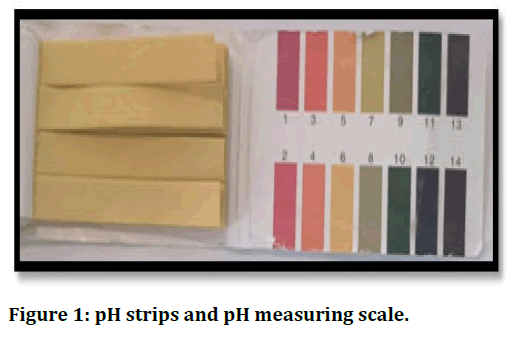
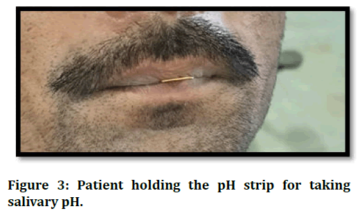
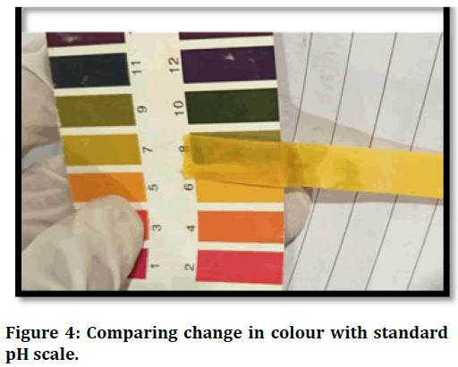
Recording salivary pH needs the collection of saliva and the pH strips. Salivary pH is measured by using pH strips between 9 am to 11 am to prevent diurnal variation. Patient was allowed to sit for 5-10 minutes in relaxed state such that saliva gets collected in the floor of mouth. A single strip was taken and kept under floor of mouth. The pH strip was removed and the change in colour should be compared with standard pH scale as shown in Figures 1-4 [13].

Figure 1: pH strips and pH measuring scale.

Figure 2: Single pH strip.

Figure 3: Patient holding the pH strip for taking salivary pH.

Figure 4: Comparing change in colour with standard pH scale.
Conclusion
The review was conducted with the purpose to evaluate the change in the salivary pH in normal healthy individuals and OSMF patients and its association with the habit. The review of literature revealed variable alterations in salivary pH. The studies demonstrated either increase, decrease or no alterations in salivary pH in given study groups. No significant alteration in baseline salivary pH is observed in normal and oral submucous fibrosis individuals. While significant alterations in post chew salivary pH were observed in normal and oral sub mucous fibrosis patient. The post chew salivary pH of oral sub mucous fibrosis individuals showed slightly alkaline pH as compared to normal individuals. The future study may be conducted with the broader representation of sample to verify the existing study finding.
References
- Maheswari TU. Assessment of salivary pH In patients with oral submucous fibrosis. Intern J Pharmaceu Res 2020; 12.
- Panchbhai A. Effect of oral submucous fibrosis on jaw dimensions. Turkish J Orthodon 2019; 32:105.
[Crossref] [Google Scholar] [PubMed]
- Lalfamkima F, Bommaji S, Reddy K, et al. Comparative evaluation of alteration in salivary flow rate between betal nut/gutkha chewers with and without OSMF and healthy subjects: A prospective case-control study. Oncol J India 2021; 5:1.
- Hande AH, Chaudhary MS, Gadbail AR, et al. Role of hypoxia in malignant transformation of oral submucous fibrosis. J Datta Meghe Instit Med Sci Univ 2018; 13:38-43.
- Donoghue M, Basandi PS, Adarsh H, et al. Habit-associated salivary pH changes in oral submucous fibrosis-A controlled cross-sectional study. J Oral Maxillofacial Pathol 2015; 19:175.
[Crossref] [Google Scholar] [PubMed]
- Shih YH, Wang TH, Shieh TM, et al. Oral submucous fibrosis: A review on etiopathogenesis, diagnosis and therapy. Intern J Molec Sci 2019; 20:2940.
[Crossref] [Google Scholar] [PubMed]
- More CB, Rao NR, Hegde R, et al. Oral submucous fibrosis in children and adolescents: Analysis of 36 cases. J Indian Soc Pedodon Prev Dent 2020; 38:190-199.
[Crossref] [Google Scholar] [PubMed]
- Shih YH, Wang TH, Shieh TM, et al. Oral submucous fibrosis: A review on etiopathogenesis, diagnosis and therapy. Intern J Molec Sci 2019; 20:2940.
[Crossref] [Google Scholar] [PubMed]
- Sarode SC, Chaudhary M, Gadbail A, et al. Dysplastic features relevant to malignant transformation in atrophic epithelium of oral submucous fibrosis: A preliminary study. J Oral Pathol Med 2018; 47:410-416.
[Crossref] [Google Scholar] [PubMed]
- Shende V, Biviji AT, Akarte N. Estimation and correlative study of salivary nitrate and nitrite in tobacco related oral squamous carcinoma and submucous fibrosis. J Oral Maxillofacial Pathol 2013; 17:381.
[Crossref] [Google Scholar] [PubMed]
- Lee JM, Garon E, Wong DT. Salivary diagnostics. Orthodon Craniofacial Res 2009; 12:206-211.
[Crossref] [Google Scholar] [PubMed]
- Humphrey SP, Williamson RT. A review of saliva: Normal composition, flow and function. J Prosthe Dent 2001; 85:162-169.
[Crossref] [Google Scholar] [PubMed]
- Tamgadge P, Wasekar R, Kulkarni S, et al. Comparative evaluation of alteration in salivary pH among gutkha chewers with and without oral submucous fibrosis and healthy subjects: A prospective case-control study. J Curr Oncol 2020; 3:8.
Author Info
Nelakurthi Vidya Maheswari* and Aarati Panchbhai
Department of Oral Medicine and Radiology, Datta Meghe Institute of Medical Sciences, Maharashtra, IndiaCitation: Nelakurthi Vidya Maheswari, Aarati Panchbhai, Salivary pH as a Diagnostic Marker in Oral Sub-Mucous Fibrosis, J Res Med Dent Sci, 2023, 11 (08): 032-035.
Received: 15-Nov-2021, Manuscript No. JRMDS-23-47370; , Pre QC No. JRMDS-23-47370 (PQ); Editor assigned: 18-Nov-2021, Pre QC No. JRMDS-23-47370 (PQ); Reviewed: 02-Dec-2021, QC No. JRMDS-23-47370; Revised: 19-Jul-2023, Manuscript No. JRMDS-23-47370 (R); Published: 16-Aug-2023
